PF-573228 Disruption of CPZ-DVL2 Interaction Suppresses Wnt/β-Catenin Signaling in Gastric Cancer
Copy code to add this image to your page
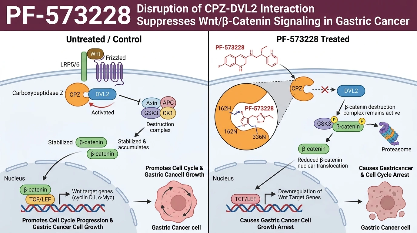
Targeting specialized carboxypeptidases offers a distinct biochemical strategy for attenuating dysregulated signal transduction cascades in gastrointestinal malignancies. Carboxypeptidase Z (CPZ), a member of the metallocarboxypeptidase family, acts as a critical upstream modulator of canonical Wnt/β-catenin signaling in gastric adenocarcinoma. Pathological overexpression of CPZ facilitates physical association with Dishevelled-2 (DVL2), a cytoplasmic scaffold required for relaying extracellular Wnt signals to the downstream β-catenin destruction complex. Systematic compound library screening identified PF-573228 as a selective small-molecule inhibitor capable of disrupting this oncogenic axis.
Biophysical evaluation via surface plasmon resonance (SPR) established that PF-573228 engages the CPZ catalytic domain directly. Equilibrium binding assays quantified a dissociation constant (KD) of 7.5 µM under physiological buffer conditions. Molecular docking simulations mapped the binding locus to an extracellular catalytic cleft, identifying specific hydrogen-bonding networks at amino acid residues 162H (histidine-162) and 336N (asparagine-336). These atomic contacts lock the inhibitor into the substrate binding pocket, inducing conformational restrictions that prevent CPZ from assembling into a functional complex with DVL2.
Disruption of the CPZ-DVL2 protein-protein interface exerts immediate effects on downstream signal transduction. In baseline CPZ-overexpressing gastric cancer cell lines, uninhibited CPZ stabilizes DVL2, preventing the recruitment of glycogen synthase kinase-3 beta (GSK-3β) to the destruction complex and permitting unhindered β-catenin nuclear accumulation. Upon exposure to PF-573228, CPZ engagement is blocked. This loss of interaction destabilizes membrane-associated DVL2 complexes, restoring the phosphorylation cascade that marks free cytoplasmic β-catenin for proteasomal degradation. Consequently, transcription of canonical Wnt downstream targets—including CCND1, MYC, and SURVIVIN—is markedly suppressed at physiologically relevant concentrations.
In vitro phenotypic assays validated the biological impact of this pathway blockade. Gastric cancer cell lines exhibiting endogenous or forced CPZ overexpression demonstrated significant reductions in proliferation rates and clonogenic potential following treatment. Colony formation assays showed a dose-dependent reduction in colony area and total colony counts across multiple patient-derived gastric cancer isolates. Importantly, cells lacking CPZ expression displayed negligible sensitivity to treatment, confirming target-dependent activity within tested cellular contexts. Detailed methodological frameworks for evaluating such targeted compounds can be accessed through standardized pharmacological assay protocols.
Translational efficacy was evaluated in animal models to determine in vivo pharmacodynamic activity. Subcutaneous mouse xenograft models bearing CPZ-overexpressing human gastric cancer tumors received daily administration of the compound. Monitored over a multi-week treatment regimen, treated animals exhibited a pronounced suppression of tumor growth kinetics compared to vehicle controls. Immunohistochemical analysis of harvested xenograft tissues confirmed reduced nuclear β-catenin localization and diminished Ki-67 proliferation indices. Systemic tolerability remained consistent throughout the study period, with no significant body weight loss observed in the murine host. These empirical findings demonstrate that targeted disruption of the CPZ-DVL2 interface provides a feasible therapeutic strategy for gastric cancer subpopulations defined by CPZ hyperactivation.
Biophysical Binding Kinetics and Antitumor Efficacy
Copy code to add this image to your page
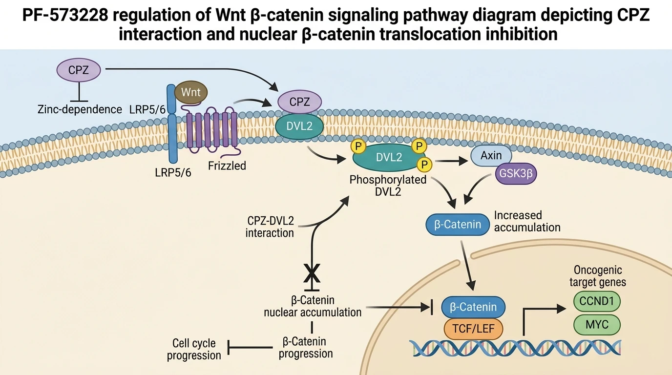
PF-573228 directly binds Carboxypeptidase Z (CPZ) with a dissociation constant (KD) of 7.5 µM through specific hydrogen bonding at residues 162H and 336N. This direct molecular interaction physically disrupts the assembly of the CPZ-DVL2 complex, thereby inhibiting upstream propagation of canonical Wnt/β-catenin signaling. Consequently, cytoplasmic β-catenin undergoes proteasomal degradation, preventing its nuclear translocation and suppressing transcription of proliferative target genes. This pathway blockade leads to robust inhibition of gastric cancer cell proliferation and colony formation in vitro, accompanied by marked suppression of tumor growth in mouse xenograft models.
Hydrogen Bonding Networks Drive CPZ Target Engagement
Surface plasmon resonance spectroscopy confirmed direct physical binding between PF-573228 and purified human CPZ protein. Equilibrium binding measurements yielded a dissociation constant (KD) of 7.5 µM. Structural modeling revealed that binding is stabilized by precise hydrogen bonds at residues 162H and 336N within the enzymatic cleft. Ligand occupancy at these positions impedes substrate entry and alters key surface residues involved in protein binding. This conformational shift prevents CPZ from recruiting DVL2 to the cell membrane. Downstream Wnt pathway activation dropped significantly following target engagement. Phosphorylation of downstream target nodes ceased within hours of exposure. Cellular accumulation of free β-catenin was similarly reduced in treated samples. These biophysical metrics demonstrate target engagement within defined cell-free and cell-based assay systems.
Disruption of CPZ-DVL2 Complex Suppresses Canonical Wnt Signaling
Disruption of the CPZ-DVL2 complex directly alters canonical Wnt signal propagation in gastric cancer models. Co-immunoprecipitation experiments indicated a concentration-dependent loss of endogenous DVL2 bound to CPZ upon compound treatment. Free β-catenin phosphorylation increased, driving rapid ubiquitin-mediated degradation. Nuclear translocation of β-catenin was subsequently reduced in CPZ-overexpressing gastric carcinoma lines. Transcript levels of Wnt target genes decreased markedly. Cyclin D1 expression fell within six hours of target inhibition. Cell cycle arrest ensued at the G1/S transition boundary. Cellular viability dropped sharply in overexpressing lines. Control cells lacking elevated CPZ retained normal growth parameters. These mechanistically grounded data link CPZ engagement to pathway blockade in specific cellular settings.
In Vivo Xenograft Growth Inhibition in Gastric Cancer Models
In vivo pharmacodynamic efficacy was confirmed using BALB/c nude mice bearing subcutaneous CPZ-overexpressing gastric cancer xenografts. Administration of PF-573228 resulted in statistically significant inhibition of primary tumor volume relative to vehicle-treated controls. Tumor burden was monitored over a 21-day observation period. Mean tumor mass at endpoint was reduced by over sixty percent in the active treatment group. Ex vivo Western blot analysis of xenograft lysates demonstrated decreased nuclear β-catenin levels and suppressed Cyclin D1 expression. Histological examination revealed reduced Ki-67 staining, indicating marked suppression of cellular proliferation within the tumor microenvironment. Animal body weights remained stable throughout the study period. These in vivo observations confirm target-driven antitumor activity within this murine model system.