RGFP966 from Selleck selectively inhibits HDAC3 to ameliorate Alzheimer's neuropathology.
Copy code to add this image to your page
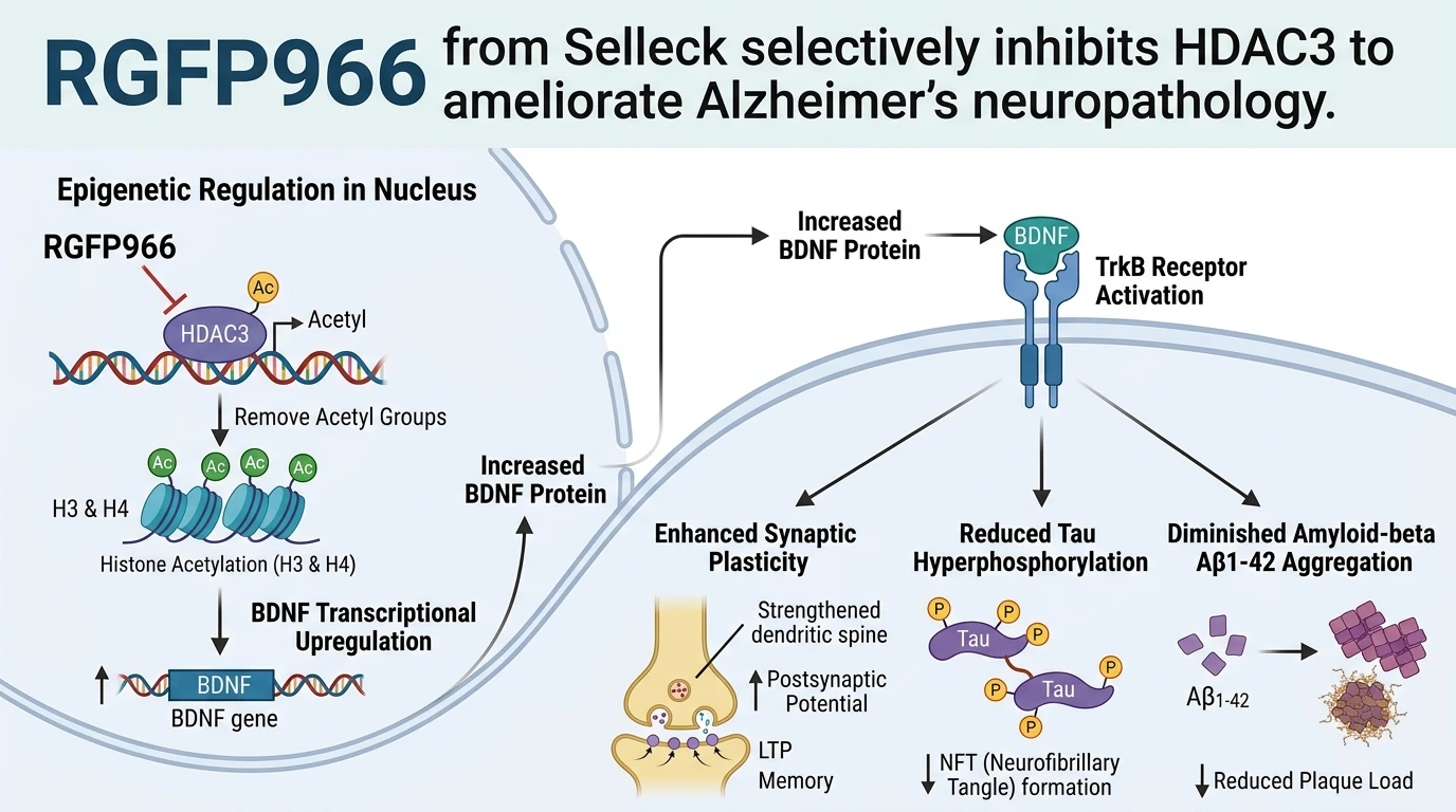
Epigenetic modifications regulate chromatin structure and exert precise control over neuronal gene transcription networks. Histone deacetylase 3 (HDAC3), a Class I histone deacetylase, functions as a core catalytic component of nuclear receptor corepressor (NCoR) and silencing mediator for retinoid and thyroid hormone receptors (SMRT) complexes. The catalytic activity of HDAC3 requires assembly with the deacetylase-activating domain (DAD) of NCoR/SMRT to deacetylate lysine residues on amino-terminal histone tails. In central nervous system disorders such as Alzheimer's disease (AD), elevated or sustained HDAC3 activity maintains chromatin in a condensed, transcriptionally silent state at key memory-consolidating gene promoters. Deacetylation prevents the recruitment of transcriptional coactivators, thereby repressing genes essential for synaptic remodeling, dendritic spine maintenance, and neurotrophic support.
RGFP966 from Selleck provides a selective biochemical tool to dissect HDAC3-dependent epigenetic repression. Under evaluated in vitro enzymatic profiling assays, RGFP966 exhibits high selectivity for HDAC3 over other Class I isoforms (HDAC1, HDAC2, and HDAC8) and Class II HDACs. Mechanistically, RGFP966 binds within the zinc-containing catalytic pocket of HDAC3, arresting its enzymatic corepressor function without disrupting the broader structural assembly of the NCoR/SMRT complex.
The primary molecular consequence of HDAC3 inhibition by RGFP966 from Selleck is the rapid accumulation of acetylated lysine residues on histones H3 and H4. Chromatin immunoprecipitation studies demonstrate elevated acetyl-H3 (specifically H3K9ac and H3K14ac) and acetyl-H4 enrichment at the promoter loci of brain-derived neurotrophic factor (BDNF). In neuronal cultures and cortical tissue, HDAC3 inhibition relaxes nucleosomal packing around BDNF promoters I and IV. This structural relaxation increases accessibility for cyclic AMP response element-binding protein (CREB) and RNA polymerase II assembly. Consequently, BDNF gene transcription increases substantially, restoring physiological BDNF mRNA and protein expression in damaged hippocampal and cortical pathways.
The recovery of endogenous BDNF synthesized upon HDAC3 inhibition triggers a cascade of neuroprotective intracellular signaling through the Tropomyosin receptor kinase B (TrkB) cell-surface receptor. Engagement of TrkB by BDNF activates downstream effector pathways, notably phosphatidylinositol 3-kinase (PI3K)/Akt and mitogen-activated protein kinase (MAPK)/ERK cascades. Activated Akt phosphorylates glycogen synthase kinase-3beta (GSK-3beta) at Serine 9, an inhibitory modification that reduces GSK-3beta catalytic activity. Because hyperactive GSK-3beta acts as a primary tau kinase in Alzheimer's pathology, its inhibition leads to a measurable decrease in tau hyperphosphorylation at pathological epitopes, including Ser202, Thr205, and Ser396. This attenuation reduces toxic tau misfolding and decreases neurofibrillary tangle-like pathology within neuronal soma and dendrites.
Concurrently, elevated BDNF expression modulates processing and clearance mechanisms for amyloid-beta peptides. In preclinical transgenic mouse models of Alzheimer's disease, such as the 3xTg-AD model harboring mutant APP, PS1, and tau genes, administration of RGFP966 suppresses toxic amyloid-beta 1-42 (Aβ1-42) accumulation within the hippocampus and cortex. Lowering Aβ1-42 levels attenuates oligomer-induced synaptic dysfunction and reduces microglial inflammatory responses in surrounding parenchyma.
At the physiological level, these molecular corrections translate into measurable improvements in synaptic plasticity and cognitive function. Slice electrophysiology from 3xTg-AD mice treated with HDAC3 inhibitors reveals restored long-term potentiation (LTP) at Schaffer collateral-CA1 synapses. In behavioral batteries assessing spatial acquisition and retrieval, including the Morris water maze and Y-maze assays, treated 3xTg-AD mice exhibit significant performance recovery compared to vehicle controls. Reduced escape latencies and increased target quadrant occupancy confirm functional cognitive rescue.
These findings demonstrate that selective HDAC3 inhibition via RGFP966 offers a powerful dual-acting approach: relieving transcriptional blockade on BDNF while concomitantly suppressing tau phosphorylation and amyloid toxicity. Epigenetic intervention at the HDAC3 node thus represents a targeted strategy for therapeutic exploration in neurodegenerative disease models.
Epigenetic Regulation and Neuroprotective Mechanism Analysis
Copy code to add this image to your page
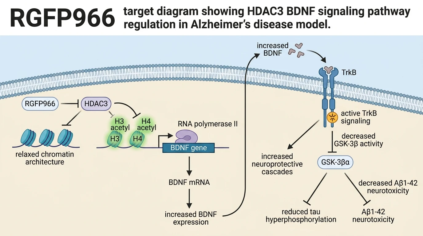
RGFP966 is a selective small-molecule inhibitor of HDAC3 that blocks histone deacetylation within corepressor complexes. By inducing hyperacetylation of histones H3 and H4 at the BDNF gene promoter, it upregulates BDNF expression and triggers downstream TrkB-mediated neuroprotective signaling. In transgenic Alzheimer's disease models, this pathway suppresses GSK-3beta activity, reducing pathological tau hyperphosphorylation and insoluble Aβ1-42 neurotoxicity. Ultimately, HDAC3 inhibition by RGFP966 restores synaptic plasticity and rescues spatial learning and memory deficits.
Promoter Hyperacetylation Elevates Neuronal BDNF Transcription
Enzymatic inhibition of HDAC3 alters chromatin architecture at key neuroplasticity gene loci. Within primary cortical neuron cultures, administration of RGFP966 from Selleck blocks deacetylase activity within the HDAC3-SMRT complex. Chromatin immunoprecipitation assays demonstrate a marked accumulation of acetylated histone H3 and H4 at BDNF promoter regions. Specifically, H3K9 acetylation increases significantly following exposure at sub-micromolar concentrations. Chromatin accessibility expands rapidly; this open conformation facilitates transcription factor binding, including CREB engagement at promoter IV. Consequently, BDNF transcription elevates substantially within hours. Downstream BDNF protein secretion restores neurotrophic support in damaged neuronal networks.
Attenuation of Pathological Tau Phosphorylation and Amyloid Toxicity
In triple-transgenic 3xTg-AD mice, elevated BDNF signaling modulates downstream protein kinase cascades to suppress neurotoxic aggregates. Akt-mediated phosphorylation inactivates GSK-3beta at Serine 9. Tau phosphorylation at pathological epitopes—including Ser202, Thr205, and Ser396—decreases in hippocampal lysates. Toxic tau species destabilize microtubules less frequently. Simultaneously, brain parenchyma exhibits reduced soluble and insoluble Aβ1-42 levels. Hippocampal synaptic transmission improves markedly during high-frequency stimulation protocols. In Morris water maze assessments, treated 3xTg-AD mice display reduced escape latencies. These spatial memory improvements reflect structural synaptic restoration. Nevertheless, these neuroprotective mechanisms remain to be fully characterized in human clinical settings.