TWS119 Suppresses Natural Killer Cytotoxicity via GSK-3β Inhibition in Breast Cancer
Copy code to add this image to your page
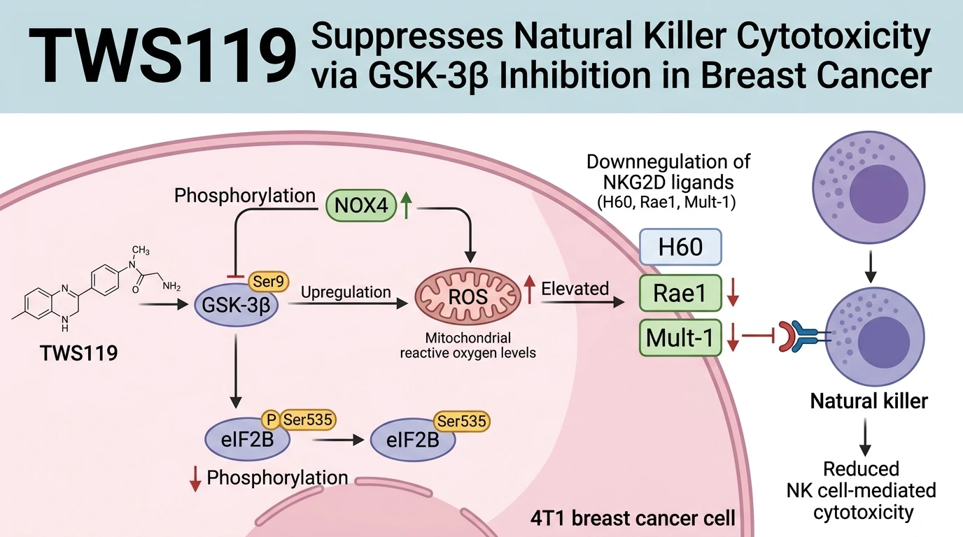
Glycogen synthase kinase 3 beta (GSK-3β) is a constitutively active serine/threonine kinase regulating diverse metabolic and immunological pathways. In murine 4T1 mammary carcinoma models, inhibitory phosphorylation of GSK-3β at Ser9 establishes a distinctive immunosuppressive cellular environment. Target engagement by TWS119, a pyrrolopyrimidine compound recognized for its GSK-3β inhibitory properties, induces Ser9 phosphorylation. Enzyme inactivation follows rapidly. This biochemical switch drives downstream metabolic restructuring inside tumor cells.
Mitochondrial homeostasis shifts significantly upon GSK-3β inhibition. NADPH oxidase 4 (NOX4) expression rises. Simultaneously, enzymatic activity within mitochondrial respiratory chain complexes I and III increases. These combined alterations elevate intracellular mitochondrial reactive oxygen species (ROS). Quantitative assays reveal sustained accumulation of superoxide radicals within treated cells. This oxidative shift alters signaling cascades critical for translational regulation.
Eukaryotic translation initiation factor 2B (eIF2B) functions as a key regulator of protein synthesis. Under physiological conditions, eIF2B phosphorylation at Ser535 modulates translation kinetics. Elevated mitochondrial ROS levels suppress this phosphorylation event. Consequently, eIF2B Ser535 phosphorylation declines markedly in 4T1 cells. This specific dephosphorylation suppresses the translation of transcripts encoding natural killer (NK) cell ligands. Surface density of NKG2D ligands drops.
Three principal NKG2D receptor ligands on murine tumor cells—H60, retinoic acid early transcript 1 (Rae1), and murine UL16-binding protein-like transcript 1 (Mult-1)—exhibit pronounced downregulation. Flow cytometric analyses confirm diminished cell-surface ligand presentation following TWS119 exposure. Lower ligand presentation compromises immune recognition. NK cells express the activating receptor NKG2D, which requires threshold ligand engagement for target cell lysis. Reduced H60, Rae1, and Mult-1 expression impairs NKG2D receptor engagement on effector NK cells. Target cell recognition fails.
Functional immunological assays demonstrate marked inhibition of NK cell-mediated cytotoxicity. Degranulation metrics, measured by surface CD107a expression, decrease significantly in NK cells co-cultured with TWS119-treated 4T1 cells. IFN-γ release by NK cells also declines. Consequently, primary tumor cells escape NK cell surveillance. In orthotopic mouse models, this loss of immune clearance correlates with accelerated primary breast tumor expansion. Pulmonary metastatic burden increases concurrently.
Reversal experiments validate the causality of this pathway. Scavenging mitochondrial ROS with targeted antioxidants restores eIF2B Ser535 phosphorylation. Restored phosphorylation restores surface expression of H60, Rae1, and Mult-1. Consequently, NK cell lytic capacity recovers fully in vitro. These experimental findings define the functional PI3K/Akt/GSK-3β/ROS/eIF2B axis in breast cancer cell immune escape.
Pharmacological Dysregulation of NK Cell Surveillance by GSK-3β
Copy code to add this image to your page
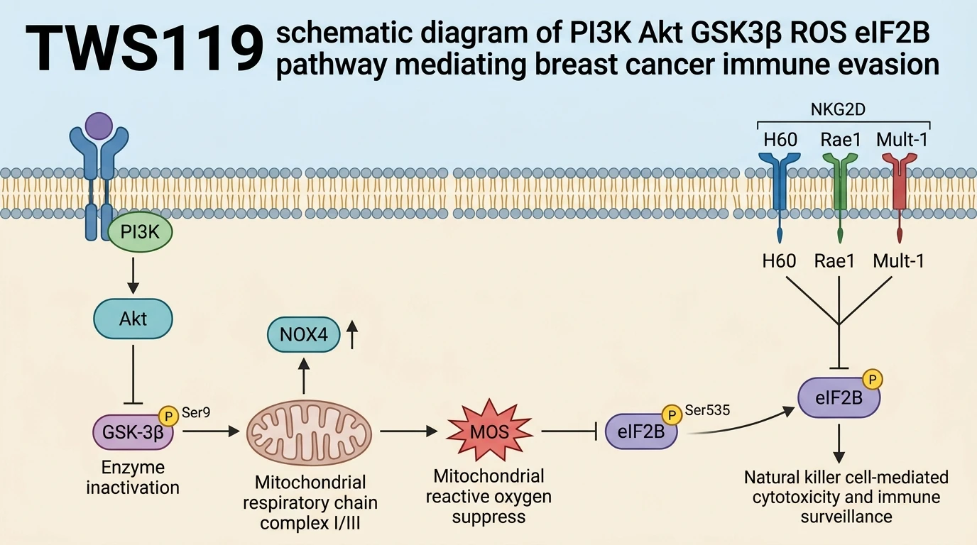
TWS119 functions as a GSK-3β inhibitor that induces enzyme inactivation via Ser9 phosphorylation. In 4T1 breast cancer models, this inactivation upregulates NOX4 and enhances mitochondrial respiratory chain complex I/III activity, generating elevated mitochondrial reactive oxygen species. High ROS levels reduce eIF2B Ser535 phosphorylation, thereby downregulating surface NKG2D ligands (H60, Rae1, Mult-1). Reduced ligand density impairs natural killer cell cytotoxicity, suppressing immune surveillance and driving tumor progression and lung metastasis. Pharmacological modulation of the PI3K/Akt/GSK-3β/ROS/eIF2B axis offers a novel pathway to restore NK cell activity.
NOX4 Upregulation and Mitochondrial ROS Accumulation in 4T1 Cells
Kinase inhibition by TWS119 alters organellar redox dynamics in 4T1 breast carcinoma cells. Ser9 phosphorylation inactivates GSK-3β, initiating transcriptional upregulation of NOX4 alongside elevated electron flux through mitochondrial respiratory chain complexes I and III. Mitochondrial ROS levels rise rapidly within hours of exposure. Elevated superoxide production acts as a secondary messenger. In vitro assays demonstrate a direct link between ROS accumulation and reduced phosphorylation of eIF2B at Ser535. This redox-sensitive modification disrupts regular translational activity without compromising overall cellular viability.
Translational Suppression of NKG2D Ligands Drives Metastatic Progression
Suppression of eIF2B Ser535 phosphorylation restricts the translation of NKG2D ligands H60, Rae1, and Mult-1. Surface expression of these key recognition molecules declines sharply on 4T1 tumor cells. Consequently, NK cell activation is severely blunted during effector-target cell interaction. In syngeneic murine models, treatment with TWS119 increases pulmonary metastasis frequency due to impaired NK cell clearance. Experimental neutralization of mitochondrial ROS reverses ligand loss and restores cytotoxic clearance. These findings highlight the therapeutic potential of targeting the PI3K/Akt/GSK-3β/ROS/eIF2B cascade in metastatic breast cancer.