Shield-1 (Shld1) Modulation of FKBP Destabilizing Domain Fusions in Membrane-Bound Enzymes
Copy code to add this image to your page
Heterologous expression systems represent indispensable tools for evaluating cytochrome P450 biotransformation kinetics and xenobiotic metabolism. In equine pharmacokinetics, cytochrome P450 3A94 (CYP3A94) functions as a key phase I biotransformation enzyme responsible for processing diverse lipophilic substrates. Optimal catalytic turnover of CYP3A94 in reconstituted cell models relies heavily on electron transfer from its obligate redox partner, NADPH P450 oxidoreductase (POR). When equine CYP3A94 is heterologously expressed in Chinese hamster lung V79 cells, basal catalytic activity toward fluorogenic probe substrates such as 7-benzyloxy-4-trifluoromethylcoumarin (BFC) remains modest due to rate-limiting endogenous electron transfer capacity. Co-expression of exogenous human POR dramatically stimulates CYP3A94 catalytic efficacy, resulting in an elevation of substrate clearance exceeding 4-fold over baseline controls.
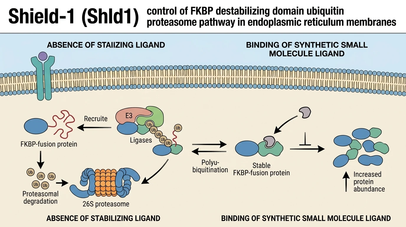
To establish precise, dynamic regulation over microsomal electron transport without altering transcription, investigators routinely implement chemical genetic tools. Among these, the FKBP-derived destabilizing domain (DD) technology offers targeted post-translational control over protein abundance. Engineered from human FKBP12 via targeted mutagenesis, the 12 kDa DD tag inherently possesses a thermally unstable, partially disordered backbone. In the unliganded state, intracellular E3 ubiquitin ligases recognize hydrophobic patches exposed on the destabilizing domain, driving rapid polyubiquitination and subsequent degradation of the target fusion protein via the 26S ubiquitin-proteasome system. Addition of the high-affinity synthetic ligand Shield-1 (Shld1) binds the mutant FKBP domain, thermally stabilizing its tertiary structure, sequestering hydrophobic domains, and inhibiting proteasomal clearance.
Application of this regulatory architecture to microsomal enzymes requires membrane tethering to the cytosolic face of the endoplasmic reticulum (ER). In an effort to tune intracellular POR activity and evaluate stoichiometric requirements for CYP3A94-mediated BFC hydroxylation, a DD tag was engineered onto the N-terminus of POR. Under standard canonical operation, omission of the small-molecule stabilizer should rapidly deplete the fusion protein, establishing a low-activity baseline state, whereas titrating the stabilizing ligand into the culture medium would predictably restore POR abundance and CYP3A94 metabolic flux in a dose-dependent manner.
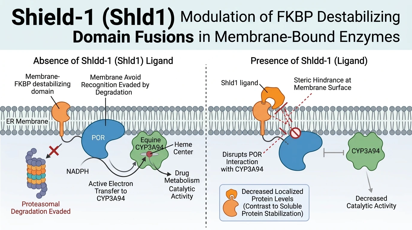
However, heterologous expression in V79 cell cultures revealed anomalous pharmacodynamic behavior for the ER-anchored DD-POR construct. In the complete absence of the ligand Shld1, the DD-POR fusion protein successfully evaded ubiquitin-proteasome system degradation. Basal expression levels of DD-POR remained persistently elevated, maintaining high catalytic throughput for equine CYP3A94 that matched levels achieved by wild-type, non-tagged POR co-expression. The destabilizing domain failed to confer the anticipated constitutive turnover in the unliganded state when fused to this integral membrane-bound electron donor.
Intriguingly, administration of Shld1 to V79 cells expressing DD-POR elicited a paradoxical inverse concentration-response profile. Increasing concentrations of Shld1 induced a progressive, dose-dependent decrease in detectable intracellular POR protein levels, which directly correlated with impaired CYP3A94 catalytic conversion of BFC. Conversely, general microsomal electron transfer capability—measured independently via extracellular cytochrome c reduction assays—remained largely unaffected by ligand exposure. These empirical findings demonstrate that while co-expressed POR is crucial for driving optimal equine CYP3A94 activity, the canonical DD stabilization model exhibits severe contextual limitations, rendering the system ineffective for post-translational expression control of ER membrane-bound enzymes such as POR.
Pharmacological Evaluation of Destabilizing Domain Kinetics
Copy code to add this image to your page
Equine CYP3A94 activity depends on co-expressed NADPH P450 oxidoreductase (POR), which increases catalytic substrate turnover over 4-fold. Attempting conditional regulation of POR using an N-terminal FKBP-derived destabilizing domain (DD) revealed unexpected failure of ligand-mediated control. In unliganded conditions, the ER-anchored DD-POR construct evaded proteasomal degradation, maintaining high basal protein and catalytic levels. Shield-1 administration paradoxically reduced POR protein levels and CYP3A94 activity in a concentration-dependent manner without altering general cytochrome c reduction. These results demonstrate that the canonical DD/Shield-1 stabilization system cannot effectively regulate membrane-bound endoplasmic reticulum proteins like POR.
POR Co-Expression Accelerates CYP3A94 Mediated Substrate Hydroxylation
Reconstitution of equine CYP3A94 activity in V79 lung fibroblasts confirmed a strong reliance on helper electron transport machinery. Baseline turnover of 7-benzyloxy-4-trifluoromethylcoumarin (BFC) remained low in cells expressing CYP3A94 alone. Transfection with human NADPH P450 oxidoreductase (POR) elevated BFC hydroxylation rates by more than four-fold. This catalytic amplification underscores the electron transfer bottleneck inherent to heterologous P450 expression systems. Direct interaction between the membrane-bound POR and CYP3A94 dictates total metabolic velocity. Steady-state kinetic assays demonstrated that maximal velocity (Vmax) increases without significant alterations in substrate binding affinity (Km). Consequently, establishing controlled stoichiometry between POR and cytochrome P450 enzymes is essential for accurate in vitro drug clearance profiling.
Anomalous Basal Stability of Endoplasmic Reticulum Anchored Fusion Constructs
To achieve tunable regulation of electron transfer, an N-terminal FKBP-derived destabilizing domain (DD) was joined to POR. In cytosolic reporter proteins, unliganded DD tags induce rapid proteasomal clearance; however, the DD-POR construct exhibited high basal stability without ligand exposure. Western blot analysis confirmed substantial accumulation of DD-POR in unsupplemented cell culture media. Correspondingly, baseline CYP3A94 activity remained fully elevated. Anchoring to the endoplasmic reticulum membrane appears to shield the destabilizing domain from cytoplasmic ubiquitin ligase complexes. Spatial orientation or steric hindrance near the lipid bilayer likely prevents hydrophobic patch recognition by quality control machinery. Consequently, the target protein evades constitutive degradation in this membrane microenvironment.
Concentration Dependent Ligand Suppression of Membrane Bound DD-POR
Subsequent addition of Shld1 yielded an unexpected pharmacodynamic response in V79 cell assays. Instead of stabilizing the fusion protein, treatment with Shld1 reduced DD-POR protein levels in a concentration-dependent manner. This loss of POR protein directly attenuated CYP3A94-mediated BFC cleavage. Notably, global microsomal electron transfer—assayed via cytochrome c reduction—showed no significant reduction following ligand exposure. The selective loss of CYP3A94 activity suggests that binding of Shld1 to the membrane-associated DD tag alters local protein-protein interactions or induces steric blockade of the P450 interaction surface. These observations indicate that the FKBP-derived destabilizing system is unsuited for regulating ER-bound enzymes like POR.