GSK-J4 Hydrochloride Histone Demethylase inhibitor Targets H3K27 Demethylase Networks in T-ALL
Copy code to add this image to your page
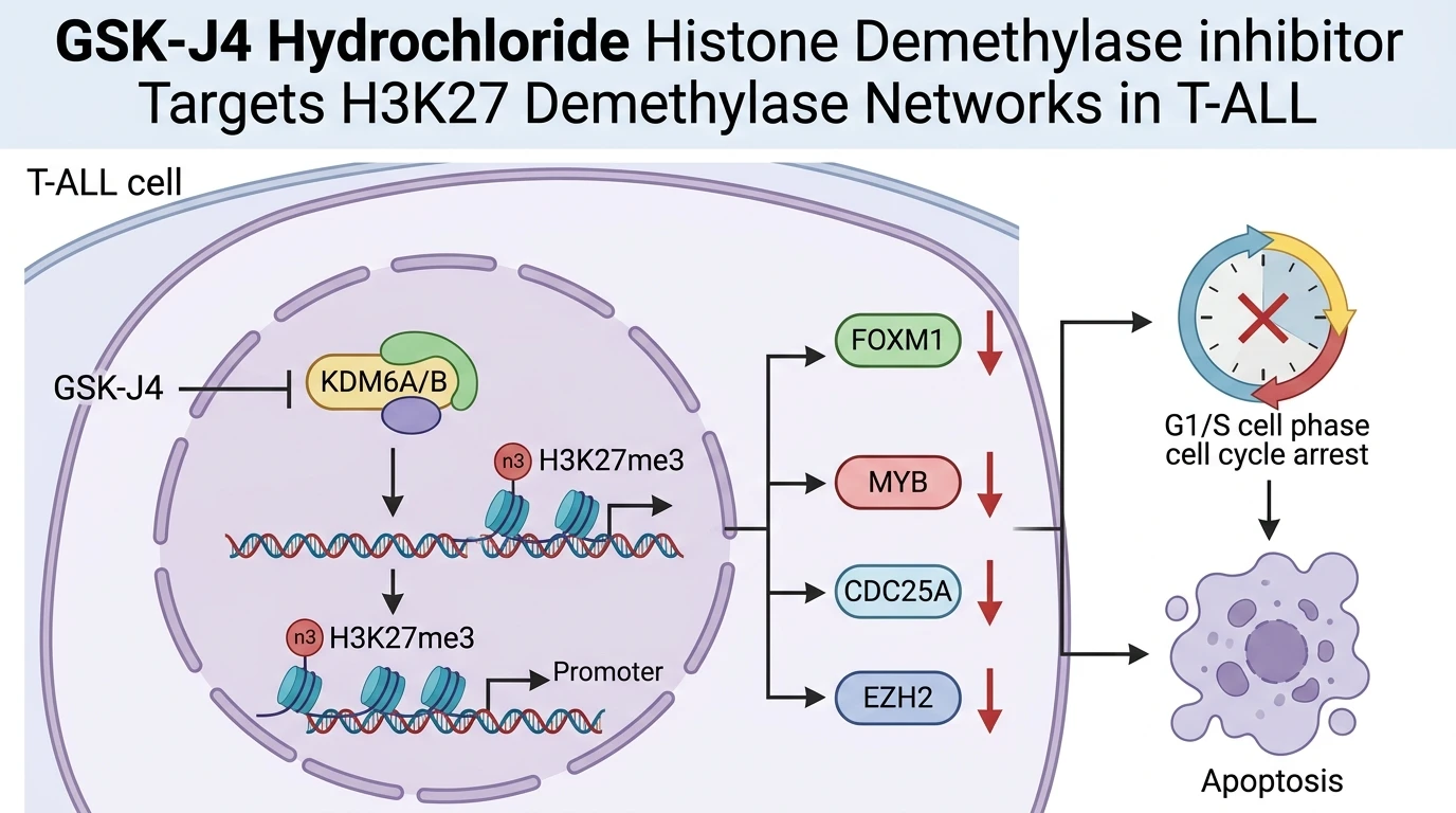
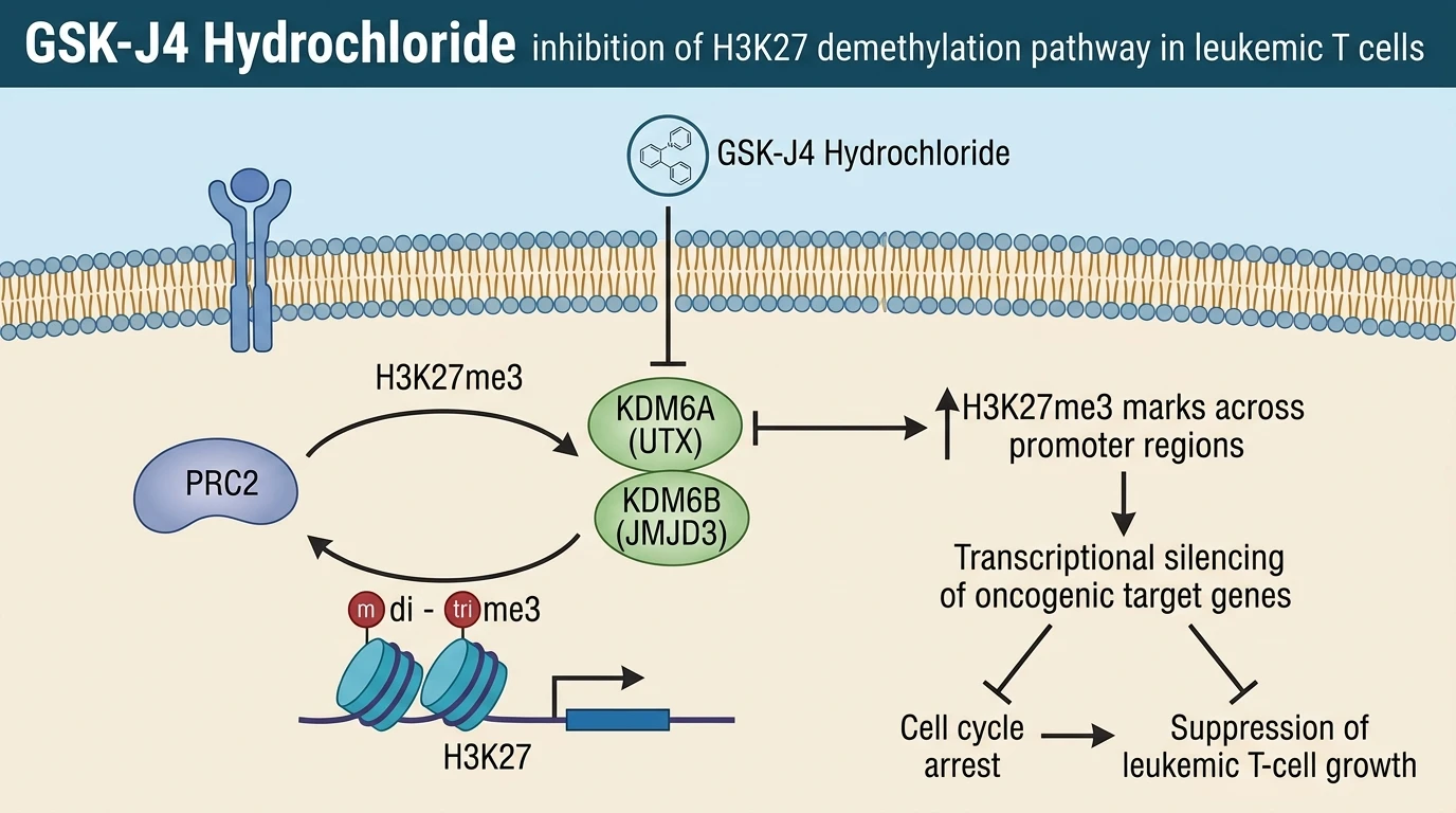
T-cell acute lymphoblastic leukemia (T-ALL) involves profound alterations in epigenetic control mechanisms. Histone H3 lysine 27 (H3K27) methylation represents a primary enzymatic determinant of chromatin condensation and transcriptional gene silencing. Catalytic removal of methyl groups from H3K27 is mediated by the KDM6 subfamily of Jumonji domain-containing Fe(II)- and 2-oxoglutarate-dependent oxygenases; specifically KDM6A (UTX) and KDM6B (JMJD3). GSK-J4 Hydrochloride functions as an ethyl ester prodrug engineered to achieve optimal intracellular uptake. Upon intracellular cleavage by non-specific esterases, the free carboxylic acid compound GSK-J1 binds the catalytic pocket of KDM6 enzymes. This active metabolite chelates the active-site iron atom and competes directly with the co-substrate 2-oxoglutarate. Catalytic inhibition was confirmed in biochemical enzyme assays; intracellular H3K27 tri-methylation (H3K27me3) levels rise rapidly following administration. Increased H3K27me3 accumulation restores local repressive chromatin marks across active promoter regions.
The crystal structure of KDM6B bound to GSK-J1 reveals specific coordination with the catalytic Fe(II) ion through a pyridine-hydrazide scaffold. This binding pocket interaction selectively blocks access of the methylated lysine substrate. In contrast, KDM4 and KDM5 demethylase family members show significantly lower binding affinity, establishing relative target selectivity for KDM6 family members under physiological buffer conditions.
Comparative transcriptomic profiling between leukemic blasts and healthy T lymphocytes reveals distinct regulatory network rewiring in T-ALL. Key transcription factors drive this altered oncogenic state. Specifically, FOXM1, MYB, and SOX4 exhibit sustained overexpression in patient-derived T-ALL samples. These transcription factors establish hyperactive regulatory circuits that sustain uncontrolled blast proliferation. Downstream effector networks further amplify these aberrant signals. MicroRNAs function as critical active nodes within these regulatory pathways; profiling identified miR-21-5p and miR-19b-3p as co-overexpressed non-coding regulators in malignant T lymphoblasts. These microRNAs directly target tumor suppressive transcripts, suppressing essential cell cycle checkpoint controls.
MYB interacts directly with promoter elements of miR-19b-3p, forming a parallel oncogenic loop that represses the PIP3 phosphatase PTEN. Loss of PTEN activity elevates signaling through the PI3K-AKT axis. SOX4 functions as a broad chromatin architect, binding super-enhancer regions that drive MYB and FOXM1 expression. These interconnected loops create an integrated survival network dependent on open chromatin states maintained by KDM6 demethylases.
The interaction between transcription factors and microRNAs generates robust feed-forward loops (FFLs). One primary circuit comprises the FOXM1-miR-21-5p-CDC25A axis. FOXM1 directly transactivates the promoter region of miR-21-5p. Simultaneously, FOXM1 and miR-21-5p synergistically upregulate CDC25A phosphatase expression. CDC25A removes inhibitory phosphate groups from cyclin-dependent kinases (CDKs), accelerating G1/S and G2/M phase transitions in lymphoblasts. Phosphatase activity remained elevated in untreated blast cultures. This dual-input mechanism buffers the cell cycle machinery against minor transcriptional fluctuations; interrupting this loop requires targeting the underlying epigenetic architecture rather than single downstream effectors.
High-throughput drug screening datasets integrated with gene expression profiles highlighted distinct epigenetic vulnerabilities in T-ALL cell models. Within these panels, the GSK-J4 Hydrochloride Histone Demethylase inhibitor demonstrated marked potency across T-ALL sub-types. Calculated IC50 metrics revealed high sensitivity in patient-derived blast cultures. Computational network analysis demonstrated strong inverse correlations between GSK-J4 sensitivity and the expression levels of core network targets. Specifically, transcription of EZH2, CDC25A, and CAPN2 declined following drug exposure. EZH2 upregulation usually compensates for altered chromatin dynamics; however, KDM6 inhibition destabilizes this cellular homeostasis.
Epigenetic intervention using GSK-J4 Hydrochloride enforces global transcriptional repatterning in T-ALL models. Accumulation of H3K27me3 at the FOXM1 and MYB promoter loci correlates directly with reduced RNA polymerase II recruitment. As FOXM1 levels decline, transcription of miR-21-5p decreases proportionally. This reduction alleviates the repression of negative cell cycle regulators while simultaneously reducing CDC25A expression. Cellular viability dropped sharply following 48 hours of treatment in vitro. Apoptosis induction was observed in primary leukemic blasts under tested assay conditions. The targeted collapse of these TF-miRNA networks demonstrates the efficacy of demethylase inhibition in controlling T-ALL cell growth.
Pharmacological Evaluation of KDM6 Demethylase Inhibition
Copy code to add this image to your page
GSK-J4 Hydrochloride acts as a cell-permeable prodrug converted to GSK-J1, which selectively inhibits KDM6 family H3K27 demethylases (KDM6A/UTX and KDM6B/JMJD3). By elevating repressive H3K27me3 marks on chromatin, the compound disrupts oncogenic feed-forward loops involving FOXM1, MYB, and miR-21-5p. This epigenetic repression downregulates key cell cycle regulators, including CDC25A and EZH2, resulting in growth arrest and apoptosis in T-ALL cells under evaluated experimental conditions.
FOXM1 and miR-21-5p Feed-Forward Loop Disruption
Targeting KDM6 demethylases directly impacts the structural accessibility of oncogenic promoters. In patient-derived T-ALL models, administration of the GSK-J4 Hydrochloride Histone Demethylase inhibitor induced a rapid increase in local H3K27me3 density at the FOXM1 promoter locus. Downstream transcription of FOXM1 ceased within 24 hours. Consequently, expression of the downstream microRNA miR-21-5p decreased significantly; this drop disrupted the FOXM1-miR-21-5p-CDC25A feed-forward loop. Cellular proliferation slowed dramatically under tested culture conditions. The G1/S phase transition was effectively blocked.
Downregulation of EZH2 and CDC25A Effector Networks
Integrated transcriptomic analyses demonstrate that KDM6 inhibition extends beyond single transcription factors. Treatment with GSK-J4 Hydrochloride caused marked reduction of CDC25A, EZH2, and CAPN2 transcripts across multiple T-ALL cell lines. CDC25A protein depletion prevented the dephosphorylation of CDK2 and CDK1; this maintained cyclin-dependent kinases in an inactive state. The selectivity margin between leukemic lymphoblasts and non-malignant T cells remained intact at sub-micromolar IC50 concentration ranges. These observations remain to be validated in randomized clinical cohorts.
Suppression of Epigenetic Plasticity in T-ALL Blasts
Epigenetic reprogramming via KDM6 family blockade enforces cell cycle arrest and primes T-ALL lymphoblasts for apoptosis. Chromatin immunoprecipitation assays confirmed widespread redistribution of H3K27me3 marks across super-enhancers controlling MYB and SOX4. The concurrent decrease in CAPN2 expression reduced calcium-dependent proteolytic signaling associated with leukemic cell survival. These preliminary data suggest that targeting H3K27 demethylation disrupts core regulatory nodes required for leukemic maintenance in this specific cellular context.