SAG (Smoothened Agonist) Visual Arrestin Mutations in Oguchi Disease Type 1
Copy code to add this image to your page
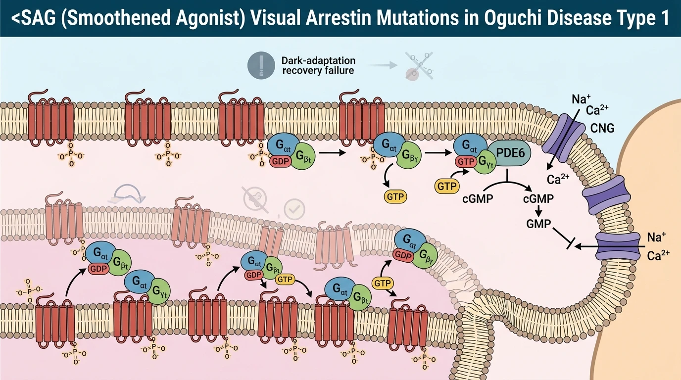
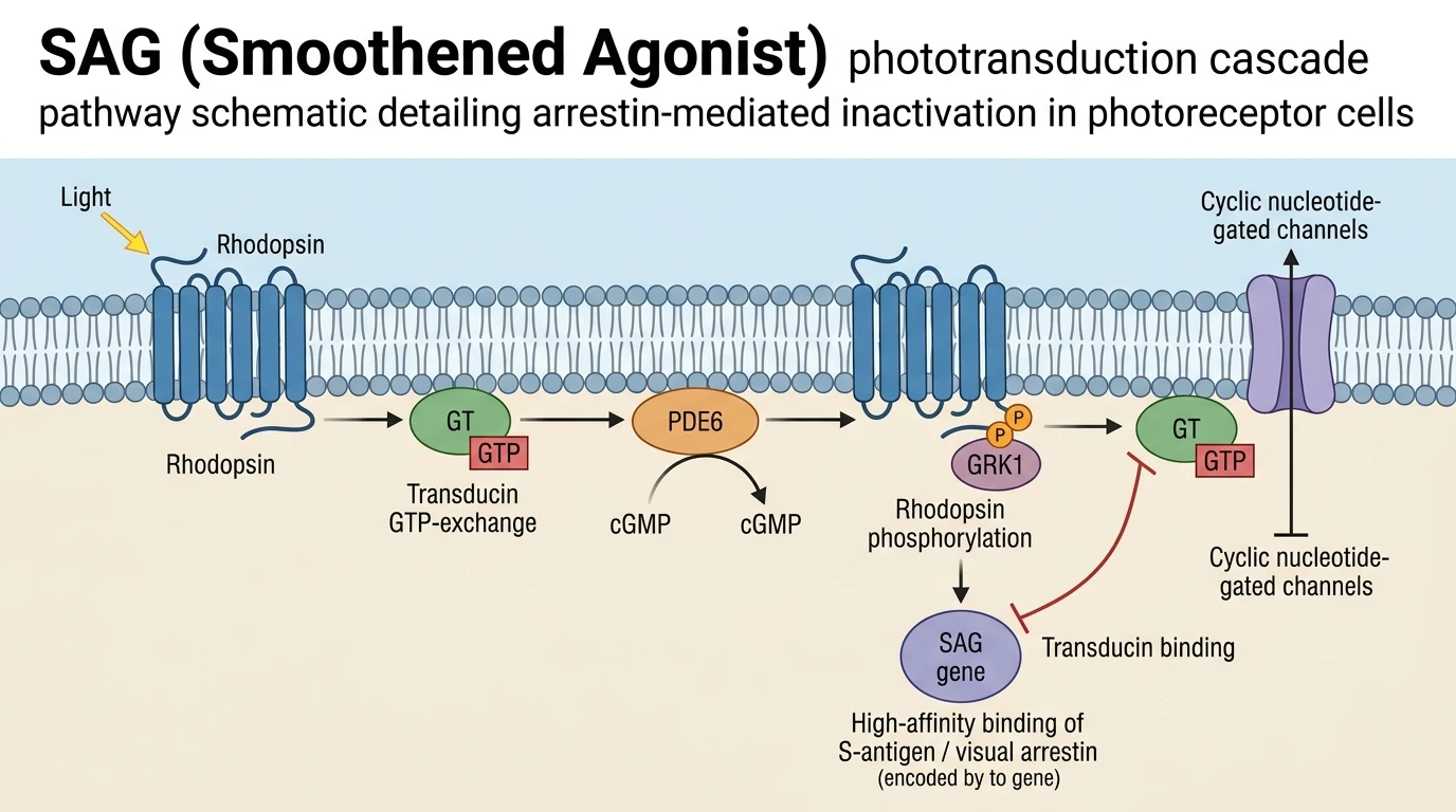
In rod photoreceptors, signal extinction following photon absorption depends on the coordinated termination of rhodopsin excitation. Visual arrestin, encoded by the SAG gene, mediates this rapid inactivation step in the phototransduction cascade pathway. Following photon capture, rhodopsin shifts to its active Metarhodopsin II conformation, catalyzing GDP-to-GTP exchange on the heterotrimeric G protein transducin. Activated transducin alpha subunits disinhibit cyclic GMP phosphodiesterase 6 (PDE6), causing rapid intracellular cGMP hydrolysis and the closure of cyclic nucleotide-gated channels. To restore dark equilibrium, rhodopsin kinase (GRK1) rapidly phosphorylates the C-terminus of active rhodopsin. Visual arrestin selectively recognizes and binds this phosphorylated, active receptor state. Binding sterically blocks further transducin engagement, halting downstream signaling.
Loss-of-function mutations within the SAG gene disrupt this homeostatic deactivation mechanism. In the absence of functional visual arrestin, activated Metarhodopsin II continues to stimulate transducin, prolonging PDE6 activation and channel closure even after light exposure ceases. This molecular persistence prevents rapid dark adaptation, giving rise to Oguchi disease type 1, a rare autosomal recessive form of congenital stationary night blindness.
Clinical and genetic investigations in two consanguineous Egyptian families established the first characterized cohorts of Oguchi disease type 1 from Egypt and the African continent. Genetic sequencing revealed two distinct homozygous pathogenic alterations within the SAG gene locus. In one family, patients carried a known homozygous nonsense mutation, p.R193* (c.577C>T). This nucleotide substitution creates a premature stop codon at position 193 of the polypeptide chain. The resulting transcript either undergoes nonsense-mediated mRNA decay or yields a truncated, unstable protein product lacking key C-terminal structural domains required for receptor binding and self-inhibition release.
In the second consanguineous Egyptian family, genetic analysis identified a novel homozygous canonical splice-site mutation, c.649-1 G>C, spanning multiple generations. The c.649-1 G>C mutation alters the highly conserved acceptor splice site preceding exon 11. In silico splicing prediction tools indicated that this substitution severely impairs wild-type splicing, inducing exon skipping or frameshift alterations that eliminate critical beta-strand frameworks within the central domain of visual arrestin. Without these structural elements, the mutant protein fails to achieve the conformation necessary to bind phosphorylated rhodopsin. For detailed biochemical structures, reference entries such as the SAG (Smoothened Agonist) compound record document target configurations across diverse signaling pathways.
Phenotypic evaluation of affected Egyptian patients demonstrated classical ocular features of Oguchi disease type 1. All affected individuals presented with lifelong, non-progressive night blindness starting in early childhood. Ophthalmoscopic examination revealed the pathognomonic Mizuo-Nakamura phenomenon. Under ambient illumination or light-adapted conditions, the ocular fundus displayed a striking diffuse golden-yellow metallic sheen. Following three hours of dark adaptation, this unusual color completely reverted to a normal reddish-pink retinal baseline. Repeat light exposure promptly restored the metallic golden discoloration. Spectral-domain optical coherence tomography (SD-OCT) provided sub-cellular structural correlates, revealing a selective loss or attenuation of the hyporeflective band corresponding to the outer segments of rod photoreceptors during light adaptation. This structural aberration reflects metabolic stress and continuous signaling downstream of unquenched rhodopsin.
Together, these genetic and phenotypic findings expand the mutation spectrum of SAG-related retinopathies. The identification of the novel canonical splice variant c.649-1 G>C confirms the role of disrupted RNA splicing in visual arrestin deficiency. Furthermore, the documented clinical features in Egyptian families establish consistency in Oguchi disease type 1 phenotypes across diverse ancestral populations, proving that visual arrestin structural integrity is indispensable for human rod photoreceptor recovery.
Genetic Disruption of Photoreceptor Recovery in Egyptian Cohorts
Copy code to add this image to your page
Loss-of-function mutations in the SAG gene disrupt the synthesis of functional visual arrestin (S-antigen). Visual arrestin normally binds phosphorylated rhodopsin to arrest transducin activation during the phototransduction cascade pathway. Without functional arrestin, activated rhodopsin signaling persists, impairing dark adaptation and photoreceptor recovery. This persistent inactivation cascade manifests phenotypically as Oguchi disease type 1, characterized by congenital stationary night blindness and the Mizuo-Nakamura phenomenon.
Homozygous p.R193* and Splice-Site Mutations Disrupt Arrestin Expression
Genetic evaluation of consanguineous Egyptian kindreds revealed two distinct homozygous loss-of-function variants within the SAG locus. In the index family, targeted sequencing identified the nonsense variant p.R193* (c.577C>T). This mutation introduces an early termination codon. Consequently, transcript degradation via nonsense-mediated mRNA decay occurs, or a truncated protein lacking crucial C-terminal domain structures is produced. In the second multi-generational family, analysis uncovered a novel canonical splice-site mutation, c.649-1 G>C. In silico splice models predicted complete loss of the consensus acceptor site. Aberrant mRNA splicing severely compromises the secondary structure of visual arrestin. Notably, research utilizing molecular probes like SAG (Smoothened Agonist) underscores how structural stability dictates pathway dynamics. In rod photoreceptors, absent visual arrestin leaves phosphorylated rhodopsin persistently active.
Clinical Manifestations and Mizuo-Nakamura Reversibility in Rod Photoreceptors
Clinical phenotyping confirmed classic features of Oguchi disease type 1 across affected individuals. Patients exhibited profound congenital night blindness under scotopic conditions. Fundus examination revealed the characteristic golden-yellow sheen. Following three hours of dark adaptation, this metallic discoloration entirely resolved—a diagnostic hallmark designated as the Mizuo-Nakamura phenomenon. Spectral-domain optical coherence tomography (SD-OCT) provided sub-cellular structural evidence. Images demonstrated attenuation of the outer segment hyporeflective band. Photoreceptor microarchitecture is thus directly impacted by prolonged signal transduction. Experimental assays using a Smoothened Agonist in hedgehog signaling demonstrate similar sensitivities to unquenched ligand activation. In visual physiology, persistent transducin activation delays intracellular ion channel restoration.
Pathogenicity Predictions and Structural Loss in Visual Arrestin Dynamics
In silico pathogenicity algorithms unanimously classified the c.649-1 G>C splice variant as deleterious. Splice site alteration disrupts the critical beta-sandwich fold required for phosphorylated rhodopsin recognition. Visual arrestin functions through a two-step activation mechanism. Phosphate moieties on the rhodopsin C-terminus release the arrestin C-tail, exposing binding surfaces for the receptor core. Without functional splice products, this regulatory cascade collapses. Rod signaling fails to reset promptly. Cone system function remains preserved, explaining intact daytime visual acuity. Expanding the genetic database of SAG mutations enhances diagnostic precision in North African populations.