TMP269 TMP269 HDAC Inhibition Profiles in Urothelial Carcinoma Models
Copy code to add this image to your page
Histone deacetylases (HDACs) regulate chromatin structure and gene expression by removing acetyl groups from lysine residues on histone tails. Among the eighteen human HDAC isoforms, class IIA enzymes—comprising HDAC4, HDAC5, HDAC7, and HDAC9—occupy a distinct biochemical niche. These proteins contain a unique N-terminal regulatory domain that mediates nucleocytoplasmic trafficking and protein-protein interactions. Small-molecule targeting of class IIA enzymes has gained interest across cardiovascular, metabolic, and neurodegenerative domains. Within oncology, evaluating selective inhibitors clarifies isoform-specific functional roles versus broad epigenetic perturbation.
TMP269 is a potent, selective inhibitor engineered to engage class IIA HDAC catalytic domains. Structurally characterized by a trifluoromethyloxadiazole (TFMO) zinc-binding moiety, TMP269 coordinates the active-site catalytic zinc ion with high selectivity over class I, class IIB, and class IV isoforms. In cell-free biochemical assays, TMP269 demonstrates nanomolar inhibitory potency. Reported half-maximal inhibitory concentrations (IC50) reach 126 nM for HDAC4, 80 nM for HDAC5, 36 nM for HDAC7, and 19 nM for HDAC9. In contrast, binding to class I enzymes (HDAC1, HDAC2, HDAC3, and HDAC8) requires sub-millimolar to micromolar thresholds. This profile provides an exceptional pharmacological window exceeding two orders of magnitude to dissect class IIA function independently of class I inhibition.
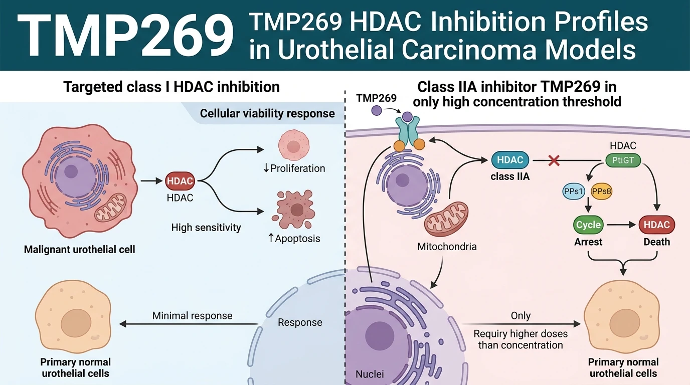
To establish therapeutic relevance in bladder malignancies, researchers evaluated TMP269 in panel assays comprising urothelial carcinoma cell lines and non-cancerous urothelial control models. In cell-free enzymatic studies, target engagement occurs at nanomolar levels. However, cell viability assays revealed a striking discrepancy. Phenotypic suppression of urothelial carcinoma growth required drug exposures exceeding 10 μM. This effective cellular threshold exceeds in vitro enzymatic IC50 values by nearly 100-fold. Such potencies suggest that isolated catalytic inhibition of class IIA HDACs does not drive antiproliferative responses in transformed urothelial cells.
Comparative analysis across non-neoplastic control cells yielded critical toxicological data. Immortalized human urothelial cells and primary normal urothelial strains displayed higher sensitivity to TMP269 than established urothelial carcinoma lines. Cell death and growth inhibition manifested at lower doses in non-cancerous cohorts. This inverted selectivity window demonstrates that class IIA catalytic function is essential for normal urothelial homeostasis rather than neoplastic persistence. Consequently, selective class IIA inhibition yields a unfavorable therapeutic index in bladder cancer models.
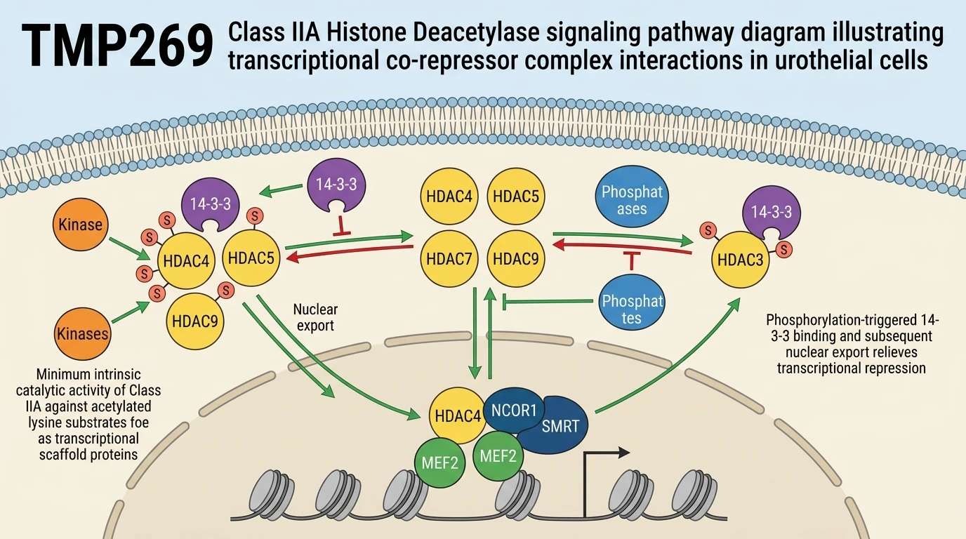
Mechanistically, these findings refine current paradigms regarding HDAC inhibitor efficacy in solid tumors. Broad-spectrum HDAC inhibitors—such as vorinostat, romidepsin, or belinostat—exhibit antiproliferative activity primarily through class I HDAC blockade. Class I isoforms (HDAC1, HDAC2, and HDAC3) display high intrinsic catalytic activity toward histone substrates; their inhibition leads to rapid histone hyperacetylation, cell cycle arrest, and apoptosis. Conversely, class IIA HDACs exhibit low catalytic activity toward native acetyl-lysine substrates due to a catalytic histidine-to-tyrosine substitution. They operate chiefly as transcriptional co-repressor scaffolds. Inhibiting the catalytic pocket with TMP269 does not displace the NCOR1/SMRT-HDAC3 corepressor assembly or alter global chromatin acetylation.
Evaluating target engagement through authoritative open-access computational resources—including chemical probe profiling databases—underscores the necessity of combining selective catalytic blockade with scaffolding disruption when studying multi-protein nuclear complexes. For urothelial carcinoma, target validation studies confirm that selective class IIA HDAC inhibition is insufficient as a standalone antineoplastic strategy.
Pharmacological Characterization and Target Validation in Urothelial Models
Copy code to add this image to your page
TMP269 achieves selective class IIA HDAC inhibition across HDAC4, HDAC5, HDAC7, and HDAC9 with nanomolar in vitro IC50 values. In urothelial carcinoma models, antiproliferative effects occur only at concentrations exceeding 10 μM—nearly 100-fold higher than cell-free enzymatic inhibition levels. Non-cancerous primary and immortalized urothelial cells exhibit greater sensitivity to TMP269 than malignant bladder lines. These observations demonstrate that catalytic inhibition of class IIA HDACs is insufficient for neoplastic suppression in urothelial carcinoma, confirming that the efficacy of broader HDAC inhibitors relies on class I isoform engagement.
Enzymatic Potency Disconnects from Cellular Proliferation Limits
Biochemical profiling confirms that TMP269 targets HDAC4, HDAC5, HDAC7, and HDAC9 at nanomolar concentrations in vitro. However, phenotypical assays in urothelial carcinoma models yield a distinct pharmacological profile. Antiproliferative activity required drug exposures above 10 μM. This concentration exceeds enzymatic IC50 values by nearly two orders of magnitude. Under standard culture conditions, low-micromolar target engagement failed to suppress cell division; phenotypic responses emerged only at high thresholds. Consequently, evaluated biochemical metrics do not translate directly into anti-neoplastic potency. Broadly targeted class I HDAC inhibitors maintain robust cytotoxic activity at sub-micromolar doses. By contrast, the selective TMP269 HDAC inhibitor profile indicates limited single-agent efficacy against transformed urothelial lineages.
Normal Urothelial Sensitivity Outpaces Transformed Cell Lines
Evaluations comparing malignant bladder lines to non-cancerous urothelial controls uncovered unexpected toxicity patterns. Primary and immortalized normal urothelial cells demonstrated heightened sensitivity to treatment. Viability in non-neoplastic controls declined at lower doses than in established carcinoma lines. Cell death in cancer models remained minimal below 10 μM—highlighting a narrow or inverted therapeutic window. These empirical findings indicate that class IIA HDAC catalytic activity is not an essential oncogenic driver in urothelial carcinoma; rather, broader pan-HDAC inhibitor efficacy stems predominantly from class I enzyme inhibition. Thus, deploying TMP269 HDAC selective agents alone does not produce an effective antitumor response in this cellular context.