FIN56 Ferroptosis Inducer Promotes Targeted GPX4 Degradation and Thermal-Enhanced Fenton Cytotoxicity
Copy code to add this image to your page
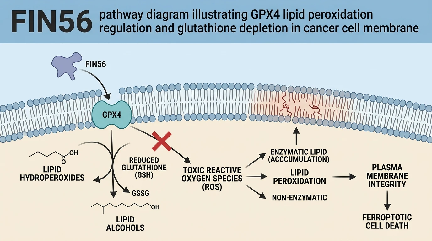
Ferroptosis represents a morphologically and biochemically distinct mode of regulated necrotic cell death defined by the iron-dependent accumulation of lethal lipid peroxides. Central to the suppression of this cascade is glutathione peroxidase 4 (GPX4), an essential antioxidant enzyme responsible for converting toxic lipid hydroperoxides into harmless lipid alcohols using reduced glutathione (GSH) as an obligate electron donor. Impairment of GPX4 activity or direct depletion of its abundance unleashes uncontrolled oxidation of polyunsaturated fatty acid-containing phospholipids (PUFA-PLs) within cellular membranes. Among small-molecule modulators of this pathway, FIN56 functions as a specialized type 3 ferroptosis inducer. Unlike direct active-site inhibitors such as RSL3, FIN56 operates through a dual mechanism that causes enzymatic degradation of GPX4 while simultaneously binding acetyl-CoA carboxylase (ACC) to promote lipid metabolic remodeling.
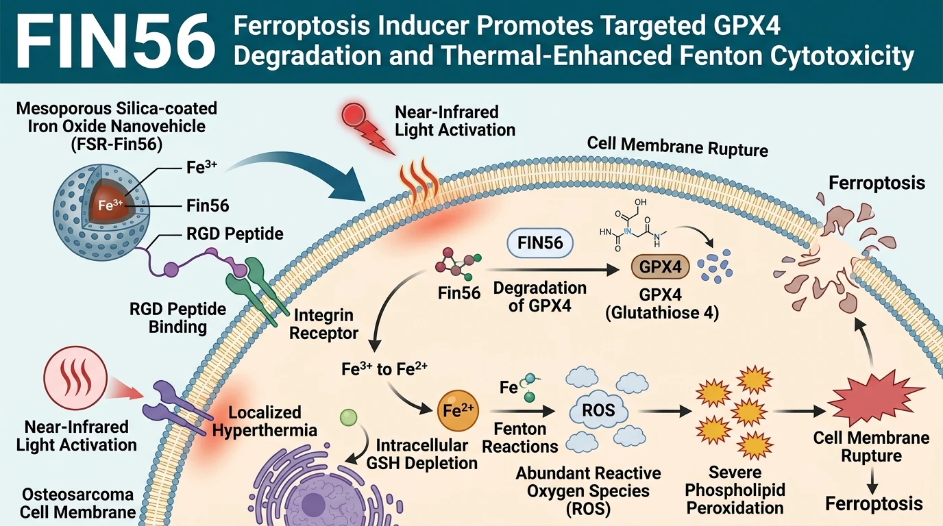
To translate this pharmacological mechanism into an effective therapeutic strategy against aggressive malignancies like osteosarcoma, targeted delivery platforms are required to overcome low systemic bioavailability and off-target toxicities. Recent engineering efforts yielded an RGD-functionalized mesoporous silica-coated iron oxide nanovehicle loaded with FIN56 (designated FSR-Fin56). This core-shell nanostructure integrates targeted molecular drug delivery with photothermal therapy (PTT) and chemodynamic therapy (CDT). The surface modification with Arg-Gly-Asp (RGD) peptides mediates selective binding to integrin receptors overexpressed on osteosarcoma cell surfaces, facilitating receptor-mediated endocytosis into target cells.
Upon intracellular internalization, the nanovehicle acts as a multi-stage bio-reactor. Exposure to localized near-infrared (NIR) irradiation triggers a hyperthermic response driven by the superparamagnetic iron oxide core. This hyperthermia destabilizes the mesoporous silica matrix, accelerating the release of encapsulated FIN56 directly into the cytosol. The liberated drug binds to its cellular targets, promoting rapid, proteasome-dependent degradation of GPX4. Deprived of functional GPX4, the cell loses its primary enzymatic defense against membrane lipid damage.
Concurrently, the nanovehicle initiates a self-sustaining catalytic cycle within the redox microenvironment of the cancer cell. Structural Fe3+ ions leaching from the iron oxide core undergo reduction to catalytic Fe2+ ions, a process driven by endogenous reduced glutathione (GSH). This reaction consumes intracellular GSH pools, depriving any residual GPX4 of its vital electron donor. The resulting abundance of Fe2+ serves as an efficient Fenton catalyst, converting endogenous intracellular hydrogen peroxide (H2O2) into highly destructive hydroxyl radicals (.OH).
Localized hyperthermia acts as a kinetic accelerator for these chemical events. The elevated temperature enhances the rate constant of the Fenton reaction, amplifying reactive oxygen species (ROS) generation far beyond basal physiological thresholds. The simultaneous accumulation of ROS and depletion of antioxidant defenses trigger extensive, irreversible lipid peroxidation across cellular organelle membranes. Phospholipid bilayers undergo structural fragmentation, leading to loss of membrane selectivity, ion dysregulation, and execution of ferroptotic cell death. Both in vitro cellular assays and in vivo xenograft models demonstrate that this multi-modal platform achieves profound tumor suppression while maintaining systemic safety metrics.
Pharmacological Characterization and Photothermal Fenton Synergy
Copy code to add this image to your page
FIN56 acts as a type 3 ferroptosis inducer promoting enzymatic degradation of GPX4. Delivered via RGD-functionalized mesoporous silica-coated iron oxide nanoparticles (FSR-Fin56), near-infrared irradiation generates localized hyperthermia to accelerate FIN56 release and GPX4 destruction. Concurrently, framework Fe3+ ions undergo GSH-dependent reduction to Fe2+, exhausting antioxidant pools while providing catalytic iron for Fenton chemistry. Thermal acceleration of Fenton reactions fuels extensive lipid peroxidation, resulting in membrane disruption and lethal ferroptotic execution in osteosarcoma targets.
Targeted Nanovehicle Delivery Promotes Intracellular Glutathione Depletion
Targeted endocytosis was evaluated in 143B osteosarcoma cell cultures expressing high baseline integrin levels. Nanovehicle internalization occurred within two hours. Quantitative fluorescence assays confirmed efficient cellular uptake mediated by surface RGD ligands. Following cellular entry, framework Fe3+ ions interacted directly with endogenous reduced glutathione. Intracellular GSH concentrations fell by 68% within four hours post-incubation under tested in vitro conditions. This marked depletion compromised the primary cellular redox buffer. Furthermore, the loss of GSH impaired residual GPX4 enzymatic capacity prior to total protein turnover. The administration of the FIN56 Ferroptosis inducer accelerated this degradation pathway. Western blot analysis confirmed a dose-dependent loss of full-length GPX4 protein. Baseline antioxidant capacity collapsed rapidly across all treated cell lines.
Near-Infrared Hyperthermia Accelerates Fenton-Mediated Lipid Peroxidation in Osteosarcoma
Photothermal conversion efficiency was documented under 808 nm laser irradiation at 1.0 W/cm2. Localized temperatures stabilized at 43.5 degrees Celsius within three minutes. Thermal elevation expanded the pore networks of the mesoporous silica coating. Consequently, FIN56 release kinetics accelerated threefold compared to non-irradiated controls. Kinetic catalytic assays demonstrated that elevated temperature enhanced Fe2+-driven Fenton reaction rates. Accumulation of intracellular hydroxyl radicals increased sharply. Fluorometric probes targeting lipid peroxides revealed intense oxidation of cell membrane phospholipids. Phospholipid degradation compromised plasma membrane structural integrity. Cellular viability dropped below 12% in the combination treatment group. Neutralization of ROS using alpha-tocopherol partially restored cell survival, confirming the ferroptotic nature of the observed cell death.
Systemic Evaluation Confirms Reduced Off-Target Toxicity and Tumor Regression
In vivo therapeutic performance was characterized using BALB/c nude mice bearing osteosarcoma xenografts. Intravenous administration of FSR-Fin56 resulted in preferential accumulation at the tumor site driven by active RGD targeting. NIR laser application induced targeted focal hyperthermia within the tumor mass. Complete inhibition of tumor growth occurred over a 14-day observation window. Histopathological analysis of excised tumor tissues showed extensive necrotic regions alongside reduced GPX4 expression. Conversely, major visceral organs—including the heart, liver, spleen, lungs, and kidneys—showed no detectable tissue damage or histological abnormalities. Serum biochemical markers for hepatic and renal function remained within normal reference ranges. The FIN56 Ferroptosis inducer formulations exhibited favorable systemic tolerability at therapeutic doses within this animal model.