GW4064 engagement of FXR represses IKBKG transcription to block cervical cancer growth
Copy code to add this image to your page
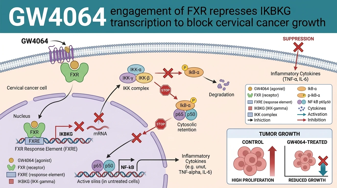
Targeting nuclear hormone receptors offers a precise strategy to intercept oncogenic transcription networks. The nuclear receptor Farnesoid X receptor (FXR; NR1H4) functions as a primary transcriptional regulator of bile acid metabolism, yet its role in gynecological malignancies remains context-dependent. Recent investigations establish that pharmacological agonist activation using targeted FXR nuclear receptor modulation directly suppresses cervical carcinoma progression through explicit transcriptional cross-talk with nuclear factor kappa B (NF-κB) cascade elements.
Endogenous FXR expression levels in human cervical cancer tissue specimens exhibit significant downregulation compared to adjacent non-tumorous cervical epithelium. Experimental siRNA-mediated knockdown of baseline FXR in non-tumorigenic cell lines leads to rapid upregulation of pro-inflammatory cytokines—including IL-6, IL-1β, and TNFα—alongside elevated basal NF-κB reporter activity. Conversely, re-establishing FXR activity using the synthetic agonist GW4064 reverses this inflammatory transcription program.
At the molecular level, activated FXR interacts directly with chromatin at specific regulatory loci. Chromatin immunoprecipitation (ChIP) assays coupled with quantitative PCR confirm that ligand-bound FXR occupies the promoter region of IKBKG (encoding inhibitor of nuclear factor kappa B kinase regulatory subunit gamma, also known as NEMO). Binding of the ligand-activated receptor complex recruits transcriptional co-repressors, selectively blocking RNA polymerase II processivity at the IKBKG locus. Consequently, intracellular levels of IKBKG mRNA and corresponding protein diminish substantially within 12 to 24 hours of treatment.
Depletion of IKBKG disrupts assembly of the core IκB kinase (IKK) complex. Without a functional IKK complex, downstream phosphorylation of IκBα at Ser32 and Ser36 fails to occur following exogenous TNFα stimulation. Unphosphorylated IκBα remains bound to p65/p50 heterodimers in the cytoplasm, preventing nuclear translocation of active NF-κB complexes. Immunofluorescence microscopy reveals that control cells display rapid p65 nuclear accumulation upon cytokine challenge, whereas treatment with GW4064 traps p65 within the cytosol.
In vitro phenotyping across HeLa (HPV-18 positive) and SiHa (HPV-16 positive) human cervical carcinoma models demonstrates dose-dependent antiproliferative responses. Real-time cell analysis and colony formation assays confirm a marked reduction in clonogenic survival following exposure to sub-micromolar concentrations of the agonist. Flow cytometry using Annexin V/Propidium Iodide staining demonstrates dose-dependent induction of apoptosis, characterized by cleavage of poly(ADP-ribose) polymerase (PARP) and caspase-3 activation. Furthermore, transwell migration and Matrigel invasion assays reflect reduced cell motility and invasive depth, corresponding with downregulation of matrix metalloproteinase-2 (MMP-2) and MMP-9 transcription.
In vivo validation utilizing BALB/c nude mice bearing subcutaneous HeLa xenografts demonstrates distinct tumor growth inhibition following daily intraperitoneal administration of the compound at 30 mg/kg. Tumor volumes in treated cohorts were reduced compared to vehicle controls over a 21-day evaluation period. Quantitative reverse transcription-PCR analysis of excised xenograft tissues showed marked reductions in IKBKG mRNA and downstream target genes, including BCL2, MMP9, and IL6. Histological evaluation of major organs (liver, kidney, heart, and spleen) revealed no treatment-related pathological alterations or serum liver enzyme elevations, confirming favorable systemic tolerance within this preclinical animal model.
To further define target selectivity, competitive binding assays confirm high-affinity engagement with the FXR ligand-binding domain (EC50 ≈ 15 nM). Reporter gene assays confirm that the observed transcriptional repression of IKBKG requires an intact FXR DNA-binding domain, as transfection with a dominant-negative FXR mutant abrogates the inhibitory effect on NF-κB activity. Furthermore, co-immunoprecipitation experiments exclude direct physical interactions between FXR and p65, reinforcing the conclusion that signal attenuation relies primarily on promoter-level regulation of the IKK regulatory subunit rather than direct protein-protein sequestering.
Pharmacological Repression of IKBKG and Tumor Inhibition
Copy code to add this image to your page
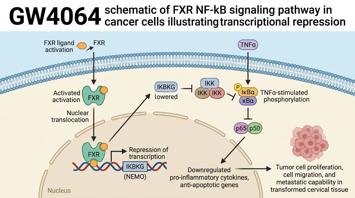
GW4064 functions as a potent agonist of the Farnesoid X receptor (FXR), inducing direct receptor binding to the promoter of IKBKG to repress its transcription. This downregulates IKBKG protein expression, preventing TNFα-induced IκBα phosphorylation and halting nuclear translocation of active NF-κB complexes. Suppressed NF-κB transcriptional activity inhibits cervical cancer cell proliferation, migration, and invasion while inducing apoptosis. In murine xenograft models, target activation significantly reduces tumor volume and pro-inflammatory cytokine expression without causing systemic organ toxicity.
Direct Promoter Binding Represses IKBKG Transcription in Vitro
Chromatin immunoprecipitation assays confirmed ligand-induced recruitment of FXR directly to promoter elements of IKBKG. Treatment with GW4064 reduced IKBKG messenger RNA expression by 65% in HeLa cells. Protein abundance fell sharply within twelve hours. Consequently, TNFα-stimulated phosphorylation of IκBα at Ser32/36 was markedly impaired under tested in vitro assay conditions. Cytosolic retention of p65 prevented nuclear NF-κB assembly; baseline transcriptional output of inflammatory cytokines dropped accordingly.
Inhibition of Cervical Cancer Cell Invasion and Apoptosis Induction
Disruption of canonical NF-κB signaling directly altered survival phenotypes in cultured SiHa and HeLa cell lines. Cellular proliferation rates decreased significantly following exposure to micro-molar concentrations of the agonist. Annexin V staining confirmed marked induction of early apoptosis; cleaved caspase-3 levels rose threefold within twenty-four hours. Transwell assays demonstrated a 50% drop in invasive cell counts—matching decreased expression of MMP2 and MMP9 transcript levels.
Xenograft Tumor Suppression Without Observed Systemic Toxicity
In vivo characterization in BALB/c nude mice bearing subcutaneous HeLa xenografts confirmed significant anti-tumor activity. Daily administration of GW4064 at 30 mg/kg suppressed mean tumor volume growth by 58% over twenty-one days. Excised xenograft tissues exhibited persistent suppression of IKBKG mRNA and reduced protein expression of pro-inflammatory markers (IL-6 and TNFα). Murine body weights remained stable throughout treatment; histological examination of liver and renal tissues showed no tissue injury or systemic necrosis.