BPTES from Selleck Targets Kidney-Type Glutaminase via Micellar Formulations for Cancer Therapy
Copy code to add this image to your page
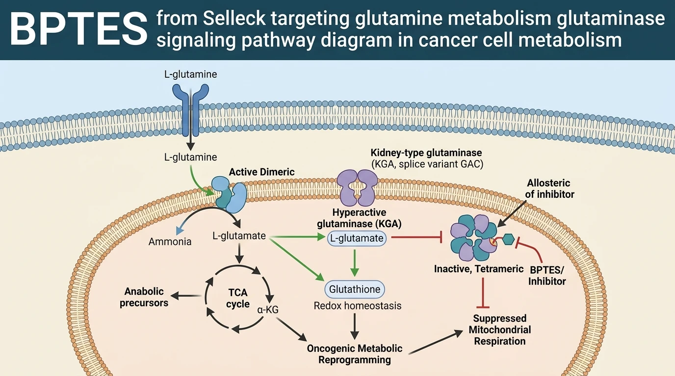
Oncogenic transformation frequently induces metabolic reprogramming, rendering cancer cells dependent on exogenous L-glutamine to sustain cellular proliferation, redox equilibrium, and biosynthesis. Kidney-type glutaminase (KGA), alongside its C-terminal splice variant glutaminase C (GAC), catalyzes the initial rate-limiting hydrolytic cleavage of L-glutamine into L-glutamate and free ammonium ions within the mitochondrial matrix. The generated glutamate serves as an essential carbon donor for alpha-ketoglutarate synthesis, directly replenishing tricarboxylic acid (TCA) cycle intermediates through anaplerosis while concurrently supporting cytosolic glutathione production. Targeted inhibition of KGA/GAC disrupts this energetic supply chain, inducing metabolic collapse in glutamine-addicted malignancies. Allosteric small-molecule inhibitors—exemplified by bis-2-(5-phenylacetamido-1,3,4-thiadiazol-2-yl)ethyl sulfide (BPTES) derivatives and 1,3,4-selenadiazole analogs such as CPD 23—bind to a hydrophobic cavity at the dimer-dimer interface of the enzyme. This allosteric engagement stabilizes a low-activity, non-catalytic tetrameric oligomer, preventing the loop movement required for active site assembly and catalytic turnover.
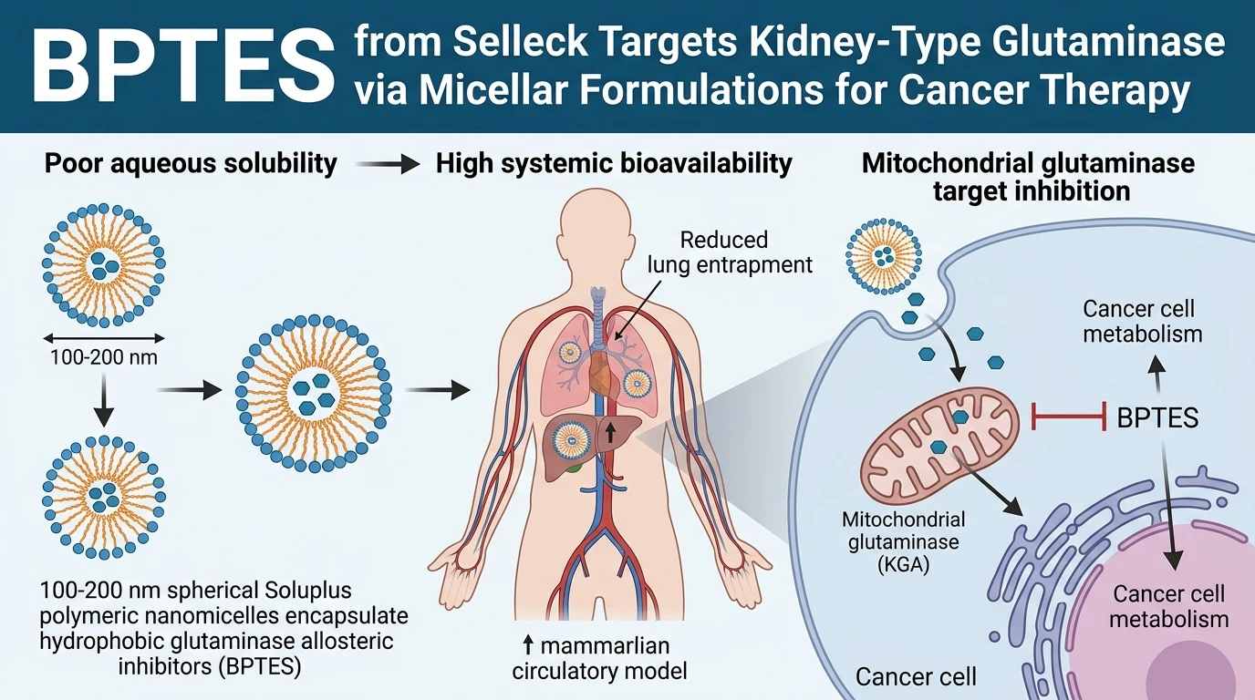
Despite high enzymatic selectivity and potent target engagement in purified recombinant assays, hydrophobic KGA allosteric inhibitors face severe biopharmaceutical limitations. High lipophilicity and poor aqueous solubility cause rapid precipitation in physiological buffers, poor oral bioavailability, and unpredictable pharmacokinetic profiles. Unmodified hydrophobic cores frequently display systemic clearance, extensive plasma protein binding, and unwanted pulmonary aggregation in animal models. To overcome these formulation bottlenecks, advanced self-assembled drug delivery platforms have been engineered to solubilize and stabilize these hydrophobic pharmacophores. In particular, a amphiphilic polyvinyl caprolactam-polyvinyl acetate-polyethylene glycol graft copolymer (Soluplus, SOL) polymeric micellar system was developed to encapsulate the 1,3,4-selenadiazole KGA inhibitor CPD 23, yielding CPD 23@SOL nanomicelles.
Physicochemical characterization demonstrated that the Soluplus polymeric micellar system increased the aqueous solubility of the hydrophobic KGA inhibitor by approximately 15,000-fold compared to unformulated free drug. Dynamic light scattering and transmission electron microscopy confirmed the spontaneous self-assembly of uniform, spherical nanoparticles spanning a diameter range of 100 to 200 nanometers. This structural dimension allows passive tumor targeting through the enhanced permeability and retention (EPR) effect while escaping rapid renal filtration. Importantly, nanomicellar encapsulation preserved the intrinsic anti-proliferative activity against target-expressing tumor cell lines, maintaining nanomolar to low-micromolar growth inhibition in vitro. When evaluated in metabolic stability assays, the micellar matrix shielded the embedded selenadiazole core from rapid phase I oxidative metabolism in liver microsomes and prevented non-specific degradation in whole blood matrices. Detailed methodological frameworks for evaluating such target-specific inhibitors can be explored through recent studies on nanomicellar formulation strategies for glutaminase inhibitors.
In vivo evaluation in rodent pharmacokinetic models demonstrated that the micellar encapsulation strategy significantly modified systemic drug disposition. Intravenous administration of CPD 23@SOL resulted in an 18-fold enhancement in relative systemic exposure (AUC) compared to free drug formulations suspended in conventional organic co-solvents. Extended circulation half-life was observed alongside reduced systemic clearance. Tissue distribution analyses revealed a striking redirection of organ accumulation: unformulated hydrophobic compound aggregates settled predominantly within pulmonary capillary beds, whereas micellar encapsulation attenuated lung retention and directed biodistribution toward hepatic tissues. This altered organotropism minimizes acute respiratory toxicity while ensuring targeted exposure to primary metabolic organs. These findings confirm that polymeric micelle encapsulation resolves critical solubility bottlenecks inherent to hydrophobic KGA allosteric inhibitors, offering a viable translational approach for glutamine-targeted cancer therapeutics.
Polymeric Encapsulation and Pharmacokinetic Disposition of Glutaminase Inhibitors
Copy code to add this image to your page
Kidney-type glutaminase (KGA/GAC) allosteric inhibitors bind the dimer-dimer interface to block glutamine hydrolysis. Soluplus polymeric micelles encapsulate hydrophobic inhibitors, increasing aqueous solubility 15,000-fold and forming stable 100–200 nm nanostructures. Micelles shield the drug from microsomal degradation, maintaining antiproliferative potency. In vivo, micellar formulations extend systemic circulation, yield an 18-fold AUC increase, and shift biodistribution from pulmonary aggregation toward hepatic accumulation.
Soluplus Nanomicelles Enhance Aqueous Solubility Fifteen Thousand Fold
Hydrophobic allosteric inhibitors of kidney-type glutaminase exhibit poor aqueous solubility. The amphiphilic polymer Soluplus self-assembles into nanomicelles with average diameters between 100 and 200 nanometers. Encapsulation enhanced aqueous solubility by 15,000-fold in ambient buffer systems. Dynamic light scattering confirmed a narrow polydispersity index and high colloidal stability over extended storage periods. The micellar hydrophobic core accommodates hydrophobic pharmacophores through non-covalent interactions. Research utilizing reference compounds like BPTES from Selleck confirms that allosteric binding pocket occupancy remains intact following micellar release. In vitro cytotoxicity assays showed that micellar encapsulation preserved low nanomolar antiproliferative efficacy against glutamine-dependent tumor cells.
Micellar Matrix Shielding Prevents Microsomal Degradation and Metabolic Clearance
Unformulated selenadiazole derivatives undergo rapid phase I metabolic degradation in hepatic fractions. Incubation with liver microsomes demonstrated that Soluplus micellar shielding prolonged half-life by protecting labile core structures from cytochrome P450 oxidation. Systemic stability in blood matrices improved significantly under physiological assay conditions. Free drug molecules precipitated rapidly upon aqueous dilution. Conversely, nanomicellar suspensions maintained structural integrity without premature payload leakage. Comparative evaluation against standard allosteric molecules, including BPTES from Selleck, verified consistent enzymatic inhibition profiles in purified GAC catalytic assays.
Eighteen Fold Exposure Enhancement Redirects Biodistribution Toward Hepatic Accumulation
In vivo pharmacokinetic trials in murine models showed an 18-fold increase in area under the plasma concentration-time curve (AUC) for encapsulated compound compared to free drug formulations. Systemic circulation time was extended substantially. Free hydrophobic inhibitors accumulated within pulmonary capillary beds, causing localized microvascular aggregation. Micellar formulation altered organ disposition dynamics markedly. Pulmonary aggregation decreased while hepatic uptake rose steadily over 24 hours. Elevated systemic exposure supported sustained target inhibition in vivo within tested xenograft models.