SP600125 suppresses JNK phosphorylation and mitigates myocardial ischemia/reperfusion apoptosis
Copy code to add this image to your page
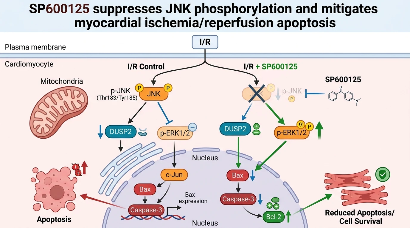
Myocardial ischemia/reperfusion injury (IRI) initiates complex biochemical cascades that induce irreversible cardiomyocyte loss. Severe oxygen deprivation during ischemia, followed by rapid reoxygenation upon reperfusion, generates elevated bursts of reactive oxygen species (ROS) and cellular calcium overload. These stress stimuli trigger activation of c-Jun N-terminal kinase (JNK), a core stress-activated protein kinase within the mitogen-activated protein kinase (MAPK) family. Upon phosphorylation, active JNK translocates to nuclear and mitochondrial compartments where it orchestrates pro-apoptotic gene expression and compromises organelle integrity. Consequently, targeted pharmacologic suppression of JNK represents a rationale-driven strategy to limit necrotic and apoptotic cell death in acute cardiac infarction models.
In experimental investigations of acute cardiac IRI, the small-molecule agent SP600125 functions as a selective reversible inhibitor of JNK activity. Application of SP600125 markedly attenuates JNK phosphorylation (p-JNK) in rodent cardiac tissues subjected to transient coronary artery occlusion. Inhibiting JNK activation prevents downstream transcription of executioner pathways responsible for cell destruction. Specifically, expression levels of the pro-apoptotic protein Bax decrease substantially following treatment with SP600125, limiting Bax transactivation and insertion into the mitochondrial outer membrane. Preserving mitochondrial integrity prevents the leakage of cytochrome c into the cytoplasm, thereby arresting the proteolytic maturation of executioner enzymes. Quantitative enzyme assays confirm a pronounced reduction in cleavage-activated caspase-3 in cardiac homogenates treated with SP600125 under reperfusion conditions.
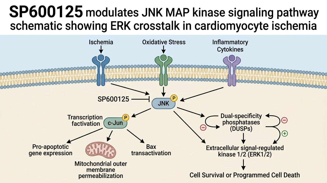
Concurrently, pharmacologic blockade of JNK by SP600125 restores anti-apoptotic signaling pathways necessary for cellular endurance. In primary neonatal rat cardiomyocytes subjected to hypoxia-reoxygenation, SP600125 elevates the expression of B-cell lymphoma 2 (Bcl-2). Raising the intracellular Bcl-2 to Bax ratio reinforces mitochondrial outer membrane resistance against opening of the mitochondrial permeability transition pore (mPTP). Beyond direct regulation of mitochondrial apoptotic proteins, JNK inhibition regulates critical crosstalk between distinct MAPK signaling networks. Under uninhibited reperfusion stress, active JNK drives expression of dual-specificity phosphatase 2 (DUSP2), a nuclear enzyme that dephosphorylates extracellular signal-regulated kinase 1/2 (ERK1/2). By blocking JNK activation, SP600125 suppresses inducible DUSP2 expression, removing a major biochemical brake on survival signaling.
Relieving DUSP2-mediated suppression allows for sustained phosphorylation of ERK1/2 (p-ERK1/2) during critical early reperfusion windows. Activated p-ERK1/2 translocates to the nucleus to induce transcription of survival genes and cardioprotective factors. Furthermore, treatment with SP600125 promotes expression of alternative regulatory phosphatases, specifically DUSP4 and DUSP16, which selectively target and limit residual JNK activity. This dynamic rebalancing of dual-specificity phosphatases establishes a self-reinforcing survival loop that favors ERK1/2 activation over pro-apoptotic JNK cascades. Notably, this biochemical sequence mirrors the cardioprotective signaling network induced by Salvianolic Acid A (SAA), a natural polyphenolic agent known to mitigate reperfusion damage. Pharmacological profile matching indicates that SP600125 reproduces the protective effects of SAA by coordinating DUSP isoform dynamics and sustaining ERK1/2 signaling.
Empirical observations across rodent models confirm that molecular inhibition translated into tangible physiological protection. Administration of SP600125 reduces myocardial infarct size as determined by triphenyltetrazolium chloride (TTC) morphometric staining following ischemic insult. Circulating levels of cardiac injury markers, including lactate dehydrogenase (LDH) and creatine kinase-MB, decline significantly in treated cohorts compared to vehicle controls. Quantitative terminal deoxynucleotidyl transferase dUTP nick end labeling (TUNEL) assays show a marked decrease in apoptotic nuclei within the ischemic border zone. Functional echocardiographic evaluations further reveal restored left ventricular ejection fraction and enhanced fractional shortening. In sum, SP600125 provides a defined mechanistic blueprint for JNK inhibition, illustrating how target-specific kinase inhibitors reorganize intracellular signaling crosstalk to preserve cardiomyocyte survival.
Pharmacological Evaluation and MAPK Crosstalk Mechanics
Copy code to add this image to your page
SP600125 acts as a selective inhibitor of c-Jun N-terminal kinase (JNK), directly inhibiting JNK phosphorylation under ischemic conditions. By suppressing JNK activity, SP600125 downregulates the expression of the inducible phosphatase DUSP2, thereby relieving catalytic inhibition on extracellular signal-regulated kinase 1/2 (ERK1/2). Sustained ERK1/2 phosphorylation promotes pro-survival transcription factors, downregulates executioner caspase-3 and pro-apoptotic Bax, and upregulates anti-apoptotic Bcl-2 along with DUSP4/16 isoforms. This dual modulation mitigates mitochondrial apoptotic signaling, curtails myocardial infarction area, and improves functional recovery following acute cardiac ischemia/reperfusion injury.
Suppression of JNK Phosphorylation Reduces Cardiomyocyte Infarct Size
JNK activity drives acute ischemic injury. In rat models of left anterior descending coronary artery occlusion, intravenous administration of SP600125 significantly attenuated myocardial infarct dimensions measured after two hours of reperfusion. Lactate dehydrogenase release decreased markedly. Western blot analysis of ventricular tissue homogenates revealed a striking reduction in phosphorylated JNK levels, accompanied by a corresponding decline in active caspase-3 cleavage products within the border zone. Cellular apoptosis dropped by over forty percent. This anti-apoptotic effect correlated with reduced pro-apoptotic Bax expression and a restored Bcl-2/Bax balance, protecting mitochondrial membrane potential under severe hypoxia.
DUSP2 Repression Restores ERK1/2 Phosphorylation and Cardioprotection
Crosstalk between MAPKs determines cell fate. Pharmacological blockade of JNK using SP600125 suppressed inducible DUSP2 phosphatase transcription in primary neonatal rat cardiomyocytes subjected to hypoxia-reoxygenation. DUSP2 down-regulation uncoupled ERK1/2 inhibition. Consequently, levels of phosphorylated ERK1/2 rose steadily during early reperfusion, driving pro-survival transcriptional programs and elevating expression of the endogenously protective phosphatases DUSP4 and DUSP16. The molecular signature mirrored Salvianolic Acid A. These findings demonstrate that targeted JNK inhibition redirects intracellular signal transduction toward pro-survival cascades, providing a mechanistic basis for cardioprotection in acute ischemic conditions.