Vismodegib (GDC-0449) targets cyclooxygenase-2 to suppress prostaglandin E2 synthesis in inflammatory models
Copy code to add this image to your page
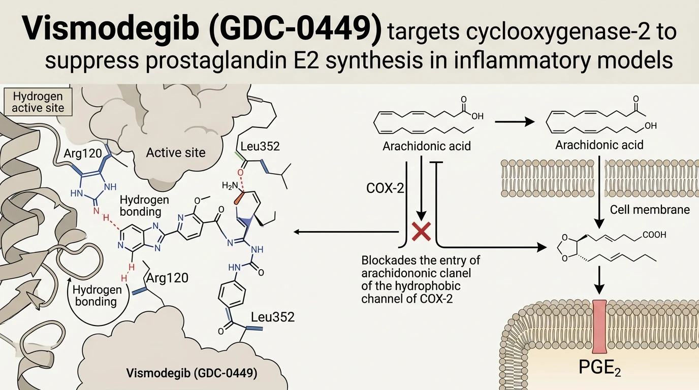
Deep-learning graph convolutional networks (GraphConvMol) trained on biophysical interaction datasets from the Directory of Useful Decoys-Enhanced (DUD-E) enable efficient repurposing screens of approved small molecules. Screening campaigns targeting human cyclooxygenase-2 (COX-2 / PTGS2) identified Vismodegib (GDC-0449), a benchmark Smoothened receptor antagonist, as a high-affinity candidate binder within the COX-2 catalytic channel. COX-2 operates as a homodimeric membrane-bound heme peroxidase that converts liberated membrane arachidonic acid into prostaglandin H2 (PGH2). Subsequent terminal synthases convert PGH2 into prostaglandin E2 (PGE2), a primary lipid mediator of acute tissue inflammation and nociception.
In silico docking analysis reveals that Vismodegib (GDC-0449) inserts directly into the primary substrate-binding hydrophobic pocket of human COX-2. Molecular dynamics simulations show distinct spatial orientation adjacent to the catalytic center. Anchoring is driven by dual hydrogen-bonding contacts with active-site residues Arg120 and Leu352. Arg120 typically coordinates the carboxylate moiety of native arachidonic acid; stable engagement of this guanidinium side chain directly obstructs fatty acid substrate entry into the hydrophobic channel. Concurrently, hydrogen bonding with the backbone nitrogen of Leu352 stabilizes the molecule within the upper binding chamber, locking the enzyme in a catalytically non-productive state. This active-site occupancy prevents iron-heme-mediated oxygenation without inducing non-specific protein denaturation under modeled thermodynamic conditions.
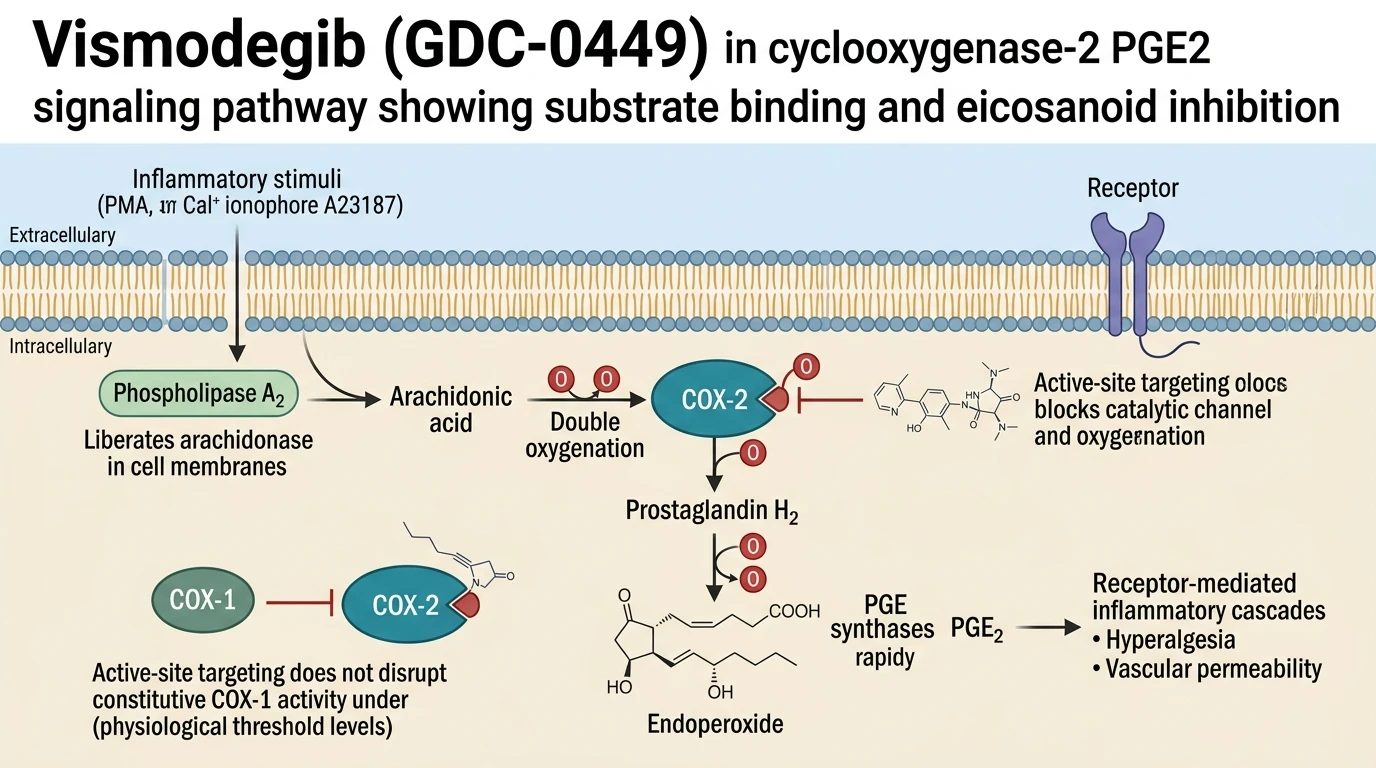
To empirically validate these computational predictions, experimental assays were performed using rat basophilic leukemia (RBL-2H3) cells. Cellular activation was induced using a combination of phorbol 12-myristate 13-acetate (PMA) and the calcium ionophore A23187. Co-stimulation with PMA and A23187 triggers robust intracellular calcium influx and protein kinase C activation, upregulating membrane phospholipase A2 cleavage and driving high-level PGE2 secretion.
When administered across a defined concentration gradient prior to stimulation, Vismodegib (GDC-0449) demonstrated significant, concentration-dependent suppression of PGE2 release into the culture supernatant. The magnitude of PGE2 inhibition produced by the compound at micromolar concentrations was statistically comparable to that observed with canonical COX-2 reference inhibitors, including celecoxib and ibuprofen. High-performance liquid chromatography and enzyme-linked immunosorbent assay (ELISA) quantifications confirmed that suppression occurred without compromising cellular membrane integrity or basal viability during the assay timeframe.
These experimental observations demonstrate that the compound possesses measurable non-canonical inhibitory activity against COX-2 in intact effector cells. The overlapping pharmacophore features that mediate Smoothened engagement appear capable of fitting the sterically restricted interior of the COX-2 cyclooxygenase active site. While further structural crystallographic verification remains necessary to confirm precise atomic coordinates, current biophysical and cell-based metrics support the classification of Vismodegib as a functional inhibitor of prostaglandin E2 biosynthesis in vitro.
Pharmacological Validation of COX-2 Active-Site Inhibition
Copy code to add this image to your page
Vismodegib (GDC-0449) inhibits cyclooxygenase-2 (COX-2) by directly binding its catalytic hydrophobic pocket. Identified via GraphConvMol deep-learning screening, the molecule forms key hydrogen bonds with active-site residues Arg120 and Leu352. This interaction blocks arachidonic acid substrate access to the catalytic center. In stimulated RBL-2H3 effector cells, the compound produces concentration-dependent suppression of prostaglandin E2 (PGE2) release, displaying potency comparable to established COX-2 inhibitors celecoxib and ibuprofen under tested in vitro assay conditions.
GraphConvMol Screening and Active-Site Hydrogen Bonding
GraphConvMol architectures process molecular topology through spatial graph representations rather than fixed fingerprint descriptors. Applying this deep-learning strategy to the DUD-E dataset revealed structural compatibility between GDC-0449 and the COX-2 catalytic pocket. In docking models, GDC-0449 occupies the hydrophobic channel. The guanidinium group of Arg120 forms a critical hydrogen bond with the compound. Simultaneously, Leu352 establishes a secondary hydrogen-bonding anchor. Substrate entry is effectively blocked. These biophysical contacts mimic classic active-site binding observed with traditional non-steroidal anti-inflammatory agents.
Concentration-Dependent Suppression of PGE2 in Cell Assays
Functional characterization required empirical testing in activated inflammatory cell lines. PMA/A23187-stimulated RBL-2H3 cells provided a robust assay system for inducible eicosanoid release. Treatment with GDC-0449 attenuated PGE2 production in a clear, dose-dependent manner under tested in vitro conditions. Efficacy levels approached those established by benchmark controls celecoxib and ibuprofen. PGE2 levels dropped sharply at low micromolar doses. Cytotoxicity assays confirmed intact plasma membranes throughout the treatment window. These findings validate the functional bioactivity of the computational hit in living cells.