iFSP1 from Selleck Inhibits FSP1 CoQ10 Reductase Activity to Overcome Ferroptosis Resistance
Copy code to add this image to your page
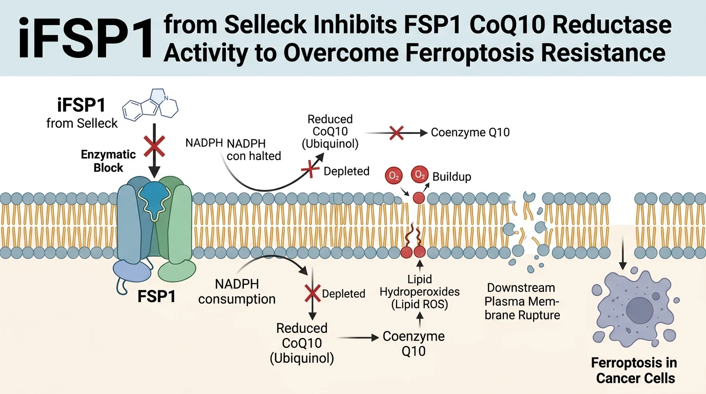
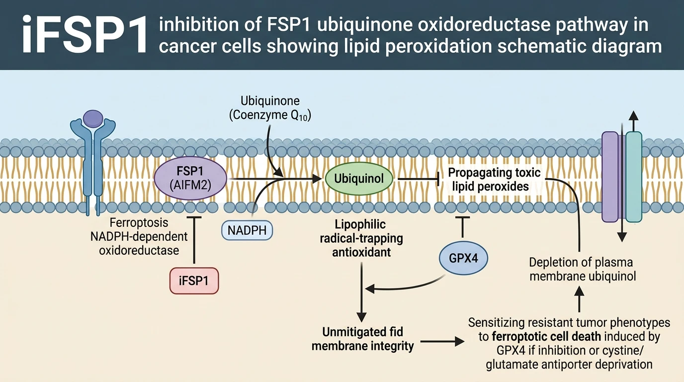
Plasma membrane lipid peroxidation represents a committed biochemical execution step during ferroptotic cell death. Ferroptosis Suppressor Protein 1 (FSP1, encoded by AIFM2) functions as an independent, non-canonical suppressor of this oxidative membrane destruction. Structurally recruited to the inner leaflet of the plasma membrane via N-terminal myristoylation, FSP1 utilizes NADPH as an electron donor to catalyze the reduction of ubiquinone (coenzyme Q10, CoQ10) into ubiquinol (CoQ10H2). Ubiquinol acts as a lipophilic radical-trapping antioxidant, directly neutralizing phospholipid hydroperoxyl radicals (L-OOH). This enzymatic axis bypasses the classic glutathione (GSH)–glutathione peroxidase 4 (GPX4) pathway. Consequently, persistent FSP1 activity confers marked resistance to canonical ferroptosis-inducing compounds, including the class I inducer erastin and the class II inducer RSL3. Pharmacological intervention targeting FSP1 catalytic flux provides a precise strategy to restore cell death sensitivity in refractory neoplastic models.
The small-molecule compound iFSP1 exhibits high biochemical selectivity for FSP1 over related oxidoreductases. Experimental characterization using iFSP1 from Selleck demonstrates competitive engagement within the catalytic binding domain of FSP1, effectively blocking its NADPH-dependent oxidoreductase function. Under controlled in vitro enzymatic assay conditions, sub-micromolar treatment with iFSP1 halts the reduction of CoQ10. This catalytic block prevents ubiquinol accumulation across the lipid bilayer. Without continuous local CoQ10H2 replenishment, lipophilic radical scavenging fails during oxidative stress. Studies in cell culture models confirm that iFSP1 does not alter total intracellular glutathione pools or decrease GPX4 protein levels. Instead, its inhibitory effect remains selectively restricted to FSP1-mediated lipid radical suppression.
Genetic and functional screens indicate that FSP1 drives ferroptosis evasion across diverse tumor backgrounds, including tuberous sclerosis complex 2 (TSC2)-deficient cells and selected ovarian and breast carcinoma lines. Research into cellular reprogramming and antioxidant resistance phenotypes highlights how metabolic alterations reinforce survival networks under stress. In these resistant configurations, elevated baseline antioxidant pathways offset lipid oxidative damage. Resistance mechanisms operate through two parallel, non-overlapping arms: the nuclear factor E2-related factor 2 (NRF2) transcriptional pathway and the FSP1 enzymatic pathway. NRF2 drives the expression of canonical antioxidant genes, including solute carrier family 7 member 11 (SLC7A11), heme oxygenase 1 (HMOX1), and ferritin heavy chain 1 (FTH1). Conversely, FSP1 functions locally at the plasma membrane to suppress lipid hydroperoxide propagation. Immunoblotting and RNA sequencing confirm that FSP1 expression is not regulated by NRF2 activity. This genetic independence demonstrates that FSP1 acts as a distinct, autonomous defense node.
Combined pharmacological inhibition assays confirm the operational redundancy of these two protective systems. In TSC2-deficient cells displaying intrinsic resistance to erastin and RSL3, treatment with iFSP1 resensitizes cells to GPX4-inhibition-induced ferroptosis. Concurrent inhibition of NRF2 using the small-molecule inhibitor ML385, combined with FSP1 suppression via iFSP1 or siRNA knockdown, completely restores ferroptosis sensitivity in vitro. Following iFSP1 administration in GPX4-inhibited cell models, lipid ROS levels—monitored via C11-BODIPY oxidation—increase rapidly within hours. Plasma membrane oxidation reaches lethal levels, triggering cell rupture. The selective restoration of ferroptotic susceptibility demonstrates that FSP1 constitutes a critical targetable survival node when canonical GPX4 activity is attenuated.
Pharmacological Evaluation and Synthetic Lethality Mechanics
Copy code to add this image to your page
iFSP1 selectively inhibits Ferroptosis Suppressor Protein 1 (FSP1/AIFM2), blocking its NADPH-dependent oxidoreductase activity at the plasma membrane. By preventing the conversion of coenzyme Q10 (ubiquinone) to its reduced radical-trapping antioxidant form (ubiquinol), iFSP1 disrupts an essential GPX4-independent antioxidant pathway. This leads to toxic accumulation of plasma membrane lipid hydroperoxides and resensitizes GPX4-inhibitor-resistant cancer cells to ferroptotic cell death.
Parallel NRF2 and FSP1 Signaling Arms Mediate Ferroptosis Resistance
Tuberous sclerosis complex 2 (TSC2) loss drives constitutive activation of mammalian target of rapamycin complex 1 (mTORC1). This metabolic shift alters cellular susceptibility to oxidative damage. In TSC2-deficient tumor models, resistance to erastin and RSL3 relies on two parallel molecular programs. The transcription factor NRF2 coordinates a broad gene expression profile. It upregulates SLC7A11 to maintain cystine import and glutathione synthesis. Concurrently, NRF2 increases HMOX1 and FTH1 levels to manage intracellular iron pools. In parallel, FSP1 acts independently at the plasma membrane. It maintains ubiquinol levels without requiring NRF2 transcriptional input. Quantitative PCR analyses show that NRF2 inhibition via ML385 leaves FSP1 transcript levels unaltered. Conversely, siRNA knockdown of FSP1 does not impair NRF2 target gene transcription. These findings establish two independent, non-overlapping branches of cellular anti-ferroptotic defense.
iFSP1 Reverses Resistance in TSC2-Deficient Ovarian and Breast Carcinomas
Pharmacological inhibition using iFSP1 from Selleck restores sensitivity to ferroptotic stimuli in refractory ovarian and breast cancer cell lines. At micromolar concentrations under standard assay conditions, iFSP1 displays minimal single-agent toxicity. However, when combined with GPX4 inhibitors like RSL3, iFSP1 triggers rapid lipid peroxidation. C11-BODIPY fluorescence oxidation increases significantly within four hours of treatment. Cellular viability drops sharply in a dose-dependent manner. Synthetic lethality occurs when both GPX4 and FSP1 enzymatic arms are disrupted simultaneously. Small-molecule suppression via ML385 further amplifies this death response in NRF2-overexpressing cell lines. These observations confirm that targeted FSP1 inhibition overcomes adaptive survival mechanisms in iron-dependent carcinoma models.
Targeting Plasma Membrane Ubiquinol Flux Resensitizes Cells to GPX4 Blockade
The primary mechanism of iFSP1 involves the selective depletion of reduced ubiquinol at the plasma membrane lipid bilayer. FSP1 normally consumes NADPH to convert ubiquinone to ubiquinol, generating a localized antioxidant shield. When iFSP1 occupies the FSP1 catalytic domain, CoQ10 reduction halts completely. Unneutralized lipid peroxides accumulate rapidly throughout the plasma membrane. In patient-derived tumor cell models, this localized disruption sensitizes resistant cells to low doses of GPX4 inhibitors. Non-transformed control cells expressing low baseline FSP1 levels maintain membrane stability during transient iFSP1 exposure. The observed therapeutic window emphasizes the context-dependent reliance of refractory cancer cells on FSP1 activity. These empirical metrics provide a biochemical foundation for dual-targeting strategies in ferroptosis-resistant tumors.